Zhang Jinde, Sun Xiang, Li Honghui, Ma Haosong, Duan Fei, Wu Zhiyou, Zhu Bowen, Chen Ronghe, Nie Liming
State Key Laboratory of Molecular Vaccinology and Molecular Diagnostics & Center for Molecular Imaging and Translational Medicine, School of Public Health, Xiamen University, Xiamen 361102 China.
Medical Research Institute, Guangdong Provincial People's Hospital, Guangdong Academy of Medical Sciences, Southern Medical University, Guangzhou 510080, China.
Photoacoustics. 2023 Feb 13;30:100462. doi: 10.1016/j.pacs.2023.100462. eCollection 2023 Apr.
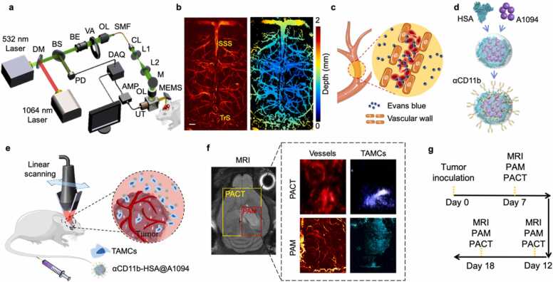
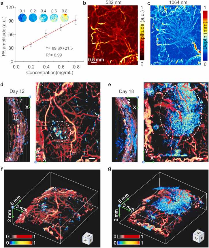
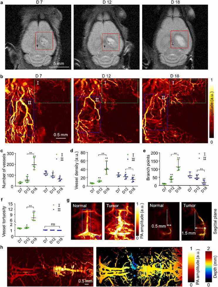
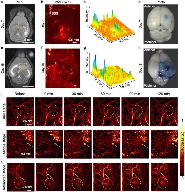
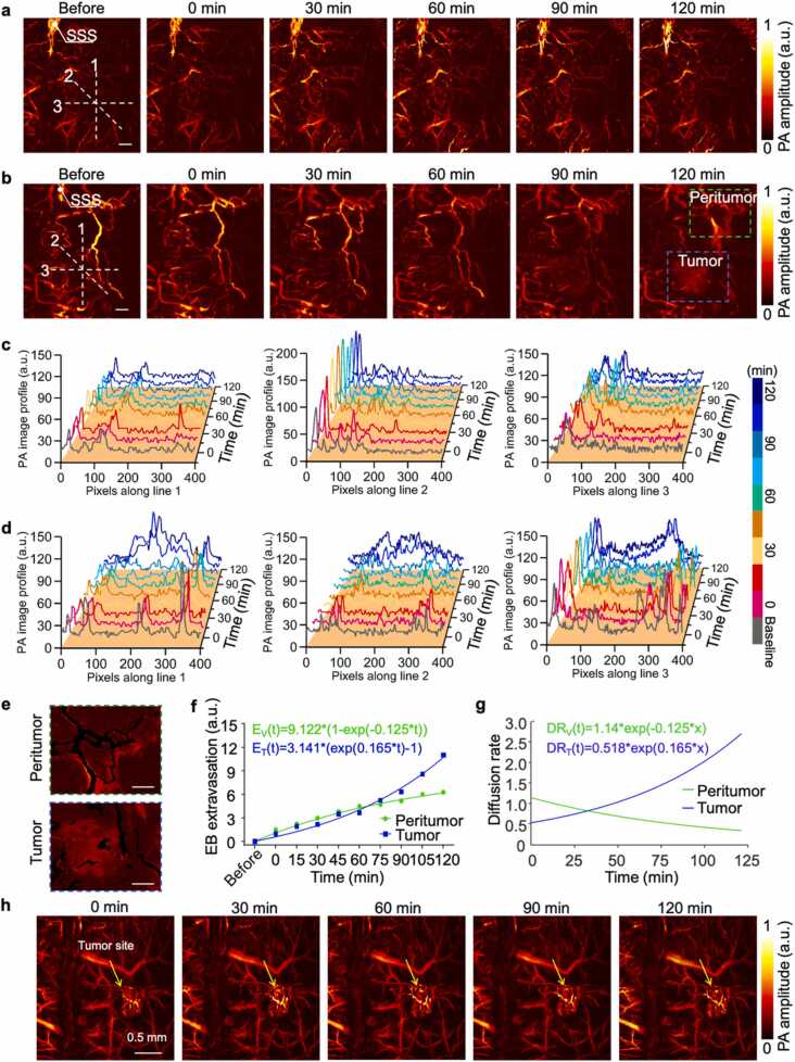
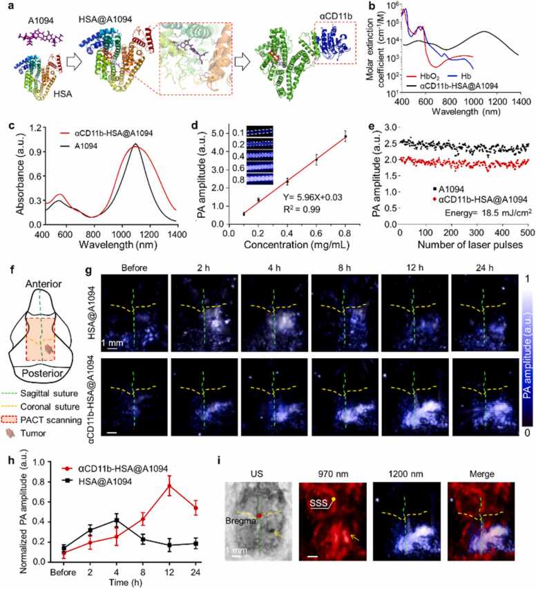
Simultaneous spatio-temporal description of tumor microvasculature, blood-brain barrier, and immune activity is pivotal to understanding the evolution mechanisms of highly aggressive glioblastoma, one of the most common primary brain tumors in adults. However, the existing intravital imaging modalities are still difficult to achieve it in one step. Here, we present a dual-scale multi-wavelength photoacoustic imaging approach cooperative with/without unique optical dyes to overcome this dilemma. Label-free photoacoustic imaging depicted the multiple heterogeneous features of neovascularization in tumor progression. In combination with classic Evans blue assay, the microelectromechanical system based photoacoustic microscopy enabled dynamic quantification of BBB dysfunction. Concurrently, using self-fabricated targeted protein probe (αCD11b-HSA@A1094) for tumor-associated myeloid cells, unparalleled imaging contrast of cells infiltration associated with tumor progression was visualized by differential photoacoustic imaging in the second near-infrared window at dual scale. Our photoacoustic imaging approach has great potential for tumor-immune microenvironment visualization to systematically reveal the tumor infiltration, heterogeneity, and metastasis in intracranial tumors.
同时对肿瘤微血管、血脑屏障和免疫活性进行时空描述,对于理解成人中最常见的原发性脑肿瘤之一——高度侵袭性胶质母细胞瘤的演变机制至关重要。然而,现有的活体成像方式仍难以一步实现这一目标。在此,我们提出一种双尺度多波长光声成像方法,该方法与独特的光学染料配合使用或不使用独特的光学染料,以克服这一困境。无标记光声成像描绘了肿瘤进展过程中新血管形成的多种异质性特征。结合经典的伊文思蓝测定法,基于微机电系统的光声显微镜能够对血脑屏障功能障碍进行动态定量。同时,使用自制的针对肿瘤相关髓样细胞的靶向蛋白探针(αCD11b-HSA@A1094),通过双尺度的第二近红外窗口的差分光声成像,可视化了与肿瘤进展相关的细胞浸润的无与伦比的成像对比度。我们的光声成像方法在肿瘤免疫微环境可视化方面具有巨大潜力,能够系统地揭示颅内肿瘤的肿瘤浸润、异质性和转移情况。