Maastricht MultiModal Molecular Imaging (M4I) Institute, Division of Imaging Mass Spectrometry, Maastricht University, Maastricht, Netherlands.
York Biomedical Research Institute, Hull York Medical School, University of York, York, United Kingdom.
Front Immunol. 2022 Jul 28;13:862104. doi: 10.3389/fimmu.2022.862104. eCollection 2022.
Spatial analysis of lipids in inflammatory microenvironments is key to understand the pathogenesis of infectious disease. Granulomatous inflammation is a hallmark of leishmaniasis and changes in host and parasite lipid metabolism have been observed at the bulk tissue level in various infection models. Here, mass spectrometry imaging (MSI) is applied to spatially map hepatic lipid composition following infection with Leishmania , an experimental mouse model of visceral leishmaniasis.
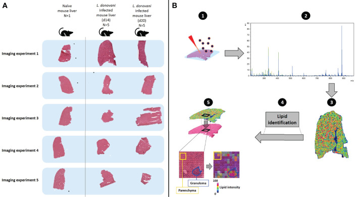
Livers from naïve and -infected C57BL/6 mice were harvested at 14- and 20-days post-infection (n=5 per time point). 12 µm transverse sections were cut and covered with norhamane, prior to lipid analysis using MALDI-MSI. MALDI-MSI was performed in negative mode on a Rapiflex (Bruker Daltonics) at 5 and 50 µm spatial resolution and data-dependent analysis (DDA) on an Orbitrap-Elite (Thermo-Scientific) at 50 µm spatial resolution for structural identification analysis of lipids.
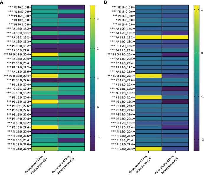
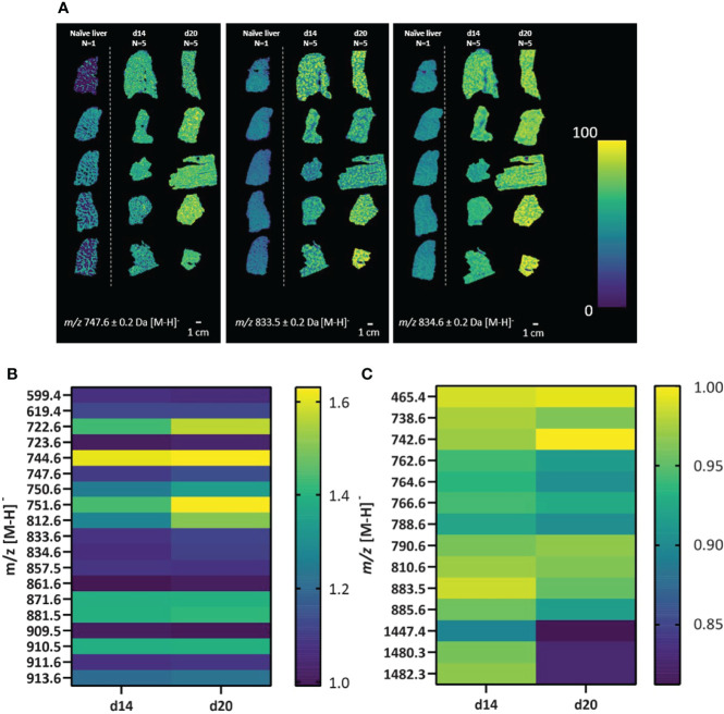
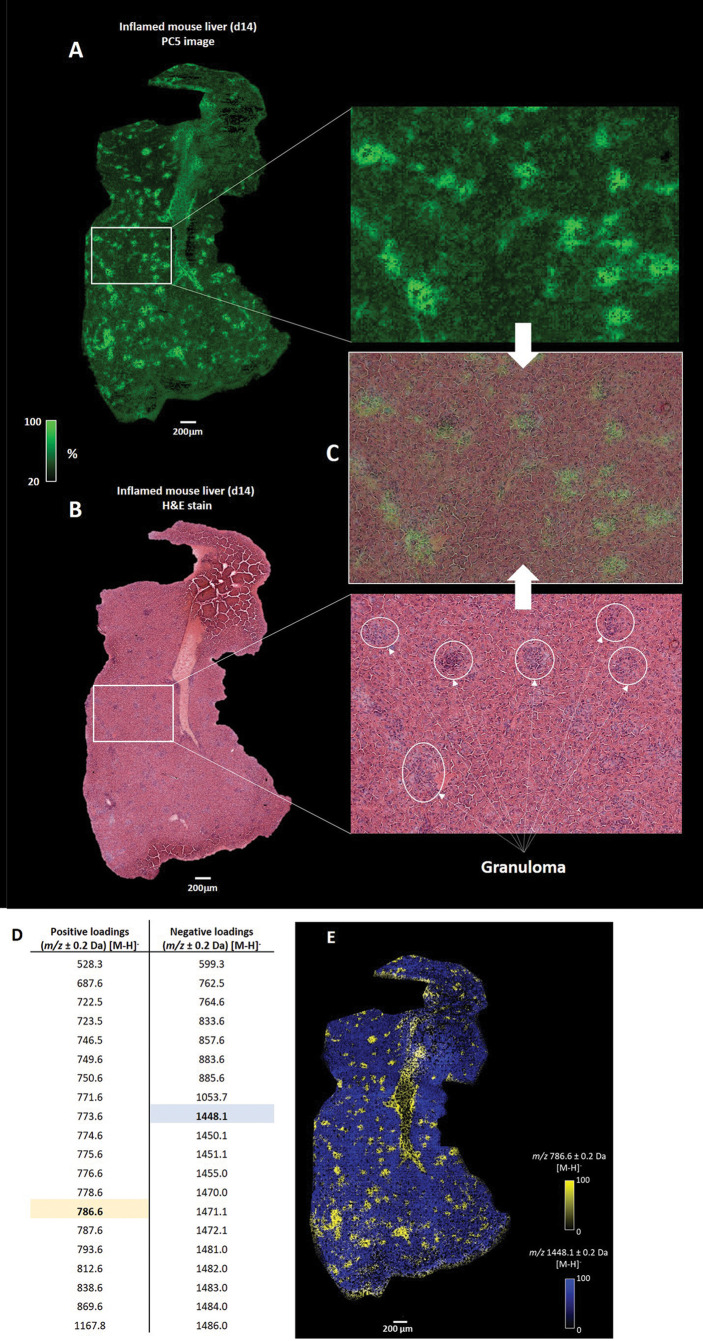
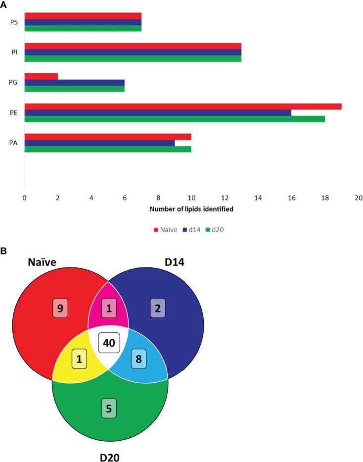
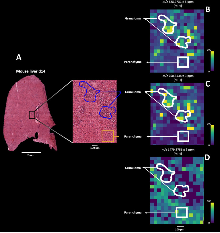
Aberrant lipid abundances were observed in a heterogeneous distribution across infected mouse livers compared to naïve mouse liver. Distinctive localized correlated lipid masses were found in granulomas and surrounding parenchymal tissue. Structural identification revealed 40 different lipids common to naïve and d14/d20 infected mouse livers, whereas 15 identified lipids were only detected in infected mouse livers. For pathology-guided MSI imaging, we deduced lipids from manually annotated granulomatous and parenchyma regions of interests (ROIs), identifying 34 lipids that showed significantly different intensities between parenchyma and granulomas across all infected livers.
Our results identify specific lipids that spatially correlate to the major histopathological feature of infection in the liver, viz. hepatic granulomas. In addition, we identified a three-fold increase in the number of unique phosphatidylglycerols (PGs) in infected liver tissue and provide direct evidence that arachidonic acid-containing phospholipids are localized with hepatic granulomas. These phospholipids may serve as important precursors for downstream oxylipin generation with consequences for the regulation of the inflammatory cascade. This study provides the first description of the use of MSI to define spatial-temporal lipid changes at local sites of infection induced by in mice.
在炎症微环境中对脂质进行空间分析是理解传染病发病机制的关键。肉芽肿性炎症是利什曼病的一个标志,在各种感染模型中,宿主和寄生虫脂质代谢的变化已在组织水平上观察到。在这里,我们应用质谱成像(MSI)来对感染后肝脏的脂质组成进行空间定位,这是一种内脏利什曼病的实验性小鼠模型。
在感染后 14 天和 20 天(n=5/时间点),从感染和未感染的 C57BL/6 小鼠中采集肝脏。将 12µm 横切片切割并覆盖 norhamane,然后使用 MALDI-MSI 进行脂质分析。MALDI-MSI 在 Rapiflex(Bruker Daltonics)上以 5 和 50 µm 的空间分辨率进行负模式,并在 Orbitrap-Elite(Thermo-Scientific)上以 50 µm 的空间分辨率进行数据依赖分析(DDA),用于脂质结构鉴定分析。
与未感染的小鼠肝脏相比,感染小鼠肝脏中脂质的丰度呈不均匀分布。在肉芽肿和周围实质组织中发现了独特的局部相关脂质质量。结构鉴定显示,40 种不同的脂质在未感染和感染的 d14/d20 小鼠肝脏中是共同的,而 15 种鉴定的脂质仅在感染的小鼠肝脏中检测到。对于病理引导的 MSI 成像,我们从手动注释的肉芽肿和实质感兴趣区域(ROI)中推断出脂质,确定了 34 种脂质在所有感染肝脏的实质和肉芽肿之间显示出显著不同的强度。
我们的结果确定了与肝脏中感染的主要组织病理学特征(即肝肉芽肿)空间相关的特定脂质。此外,我们发现感染肝脏组织中磷脂酰甘油(PG)的数量增加了三倍,并提供了直接证据表明含有花生四烯酸的磷脂定位于肝肉芽肿。这些磷脂可能作为下游氧代脂质生成的重要前体,对炎症级联的调节产生影响。本研究首次描述了使用 MSI 来定义感染引起的小鼠局部感染部位的时空脂质变化。