Rudnicki Wojciech, Piegza Tomasz, Rozum-Liszewska Natalia, Górski Mateusz, Popiela Tadeusz J, Basta Pawel, Heinze Sylwia, Luczynska Elzbieta
Electroradiology Department, Faculty of Health Sciences, Jagiellonian University Collegium Medicum, Krakow, Poland.
Maria Sklodowska-Curie National Research Institute of Oncology in Cracow, Poland.
Pol J Radiol. 2021 Mar 15;86:e159-e164. doi: 10.5114/pjr.2021.104834. eCollection 2021.
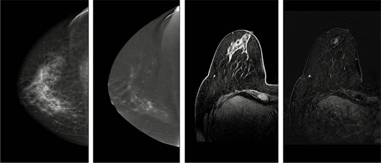
Breast cancer is the most common cause of death from neoplastic disease in women. Among all breast anatomy types, glandular type is the most problematic concerning evaluation. While digital mammography still remains the basic diagnostic tool, one must be aware of its limitations in dense breasts. Although magnetic resonance imaging (MRI) has greatly improved sensitivity, its specificity is low. Moreover, there are contraindications for MRI for some patients, so a substitute has been searched for. This study was performed to check if contrast-enhanced spectral mammography (CESM) can be a viable option for patients with dense breasts.
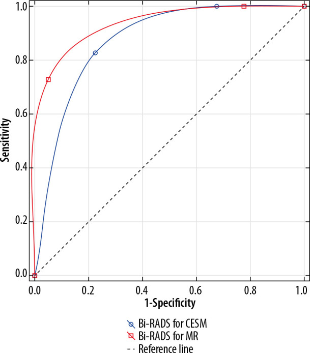
The study involved 121 patients with abnormalities detected on base-line diagnostic imaging (ultrasound or mammography). The patients had subsequent examinations, both CESM and MRI performed within a maximum 2-month time interval. The sensitivity and specificity of both methods in the whole group as well as in specific breast structure types were measured and compared.
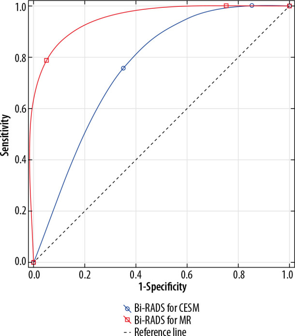
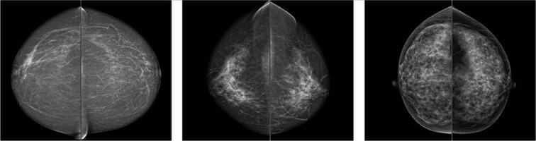
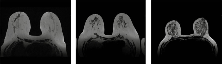
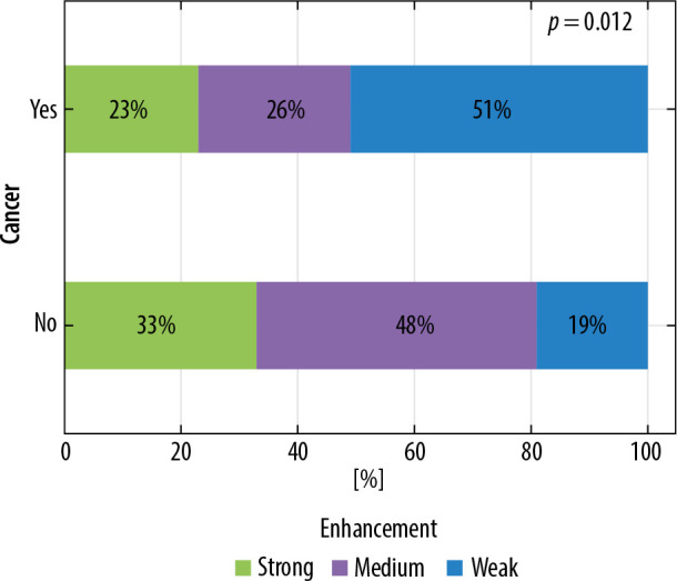
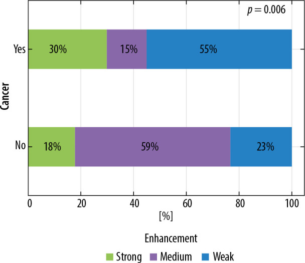
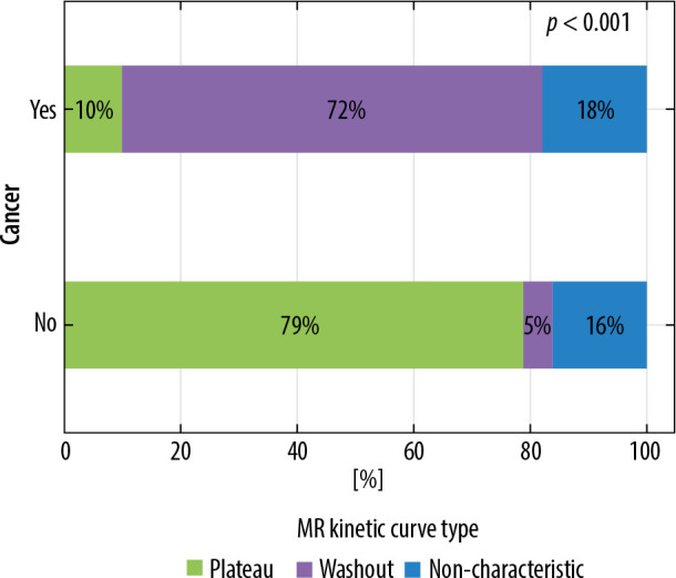
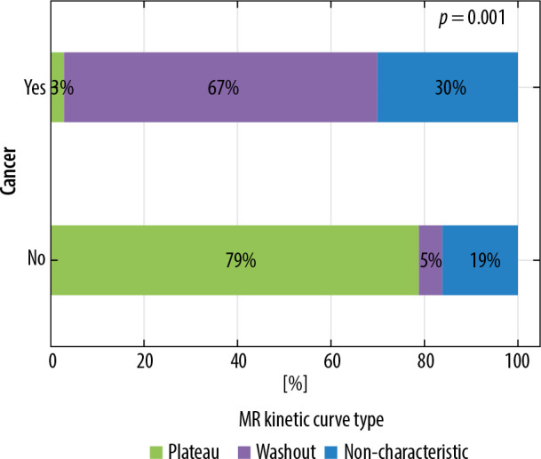
Contrast enhancement was visible in all 121 cases on MRI, while on CESM lack of enhancement was noted in 13 cases. All of those 13 lesions turned out to be benign. There were 40 (33%) benign and 81 (69%) malignant tumours. The analysed group included 53 (44%) glandular type breast patients, 39 (32%) mixed type, and 29 (23%) fatty type. Although MRI proved to be slightly more effective in dense breasts, both methods showed similar results in the whole study group.
CESM can be used with confidence in patients with glandular breast type when MRI is not available or there are reported contraindications to MRI.
乳腺癌是女性肿瘤性疾病致死的最常见原因。在所有乳腺解剖类型中,腺体型在评估方面问题最大。虽然数字乳腺摄影仍是基本的诊断工具,但必须意识到其在致密型乳腺中的局限性。尽管磁共振成像(MRI)极大地提高了敏感性,但其特异性较低。此外,部分患者存在MRI检查的禁忌证,因此一直在寻找替代方法。本研究旨在检验对比增强光谱乳腺摄影(CESM)对于致密型乳腺患者是否是一种可行的选择。
本研究纳入了121例在基线诊断成像(超声或乳腺摄影)中发现异常的患者。这些患者随后接受了检查,CESM和MRI均在最长2个月的时间间隔内进行。测量并比较了两种方法在整个研究组以及特定乳腺结构类型中的敏感性和特异性。
121例患者的MRI检查均可见对比增强,而CESM检查中有13例未见增强。这13个病灶均为良性。共有40个(33%)良性肿瘤和81个(69%)恶性肿瘤。分析的研究组包括53例(44%)腺体型乳腺患者、39例(32%)混合型患者和29例(23%)脂肪型患者。虽然MRI在致密型乳腺中显示出略高的有效性,但两种方法在整个研究组中结果相似。
当无法进行MRI检查或有MRI检查禁忌证报告时,CESM可放心用于腺体型乳腺患者。