Solano Mendoza Patricia, Aceytuno Poch Paula, Solano Reina Enrique, Solano Mendoza Beatriz
Department of Orthodontics and Dentofacial Orthopedics, School of Dentistry, University of Seville, 41009 Sevilla, Spain.
J Clin Med. 2022 Aug 9;11(16):4652. doi: 10.3390/jcm11164652.
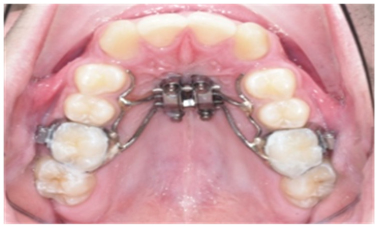
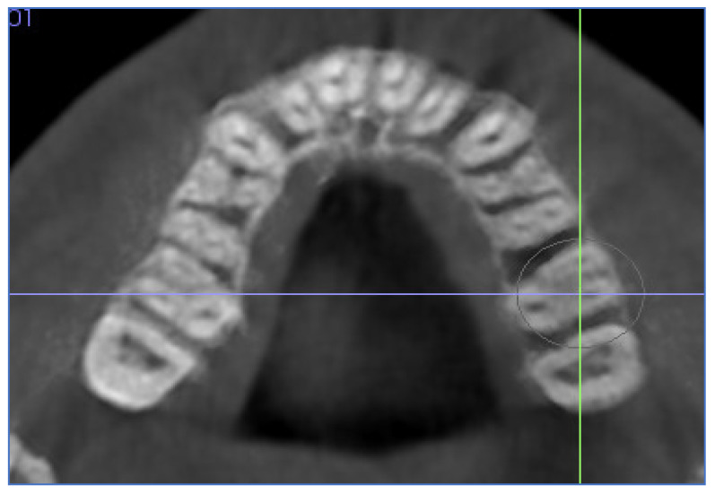
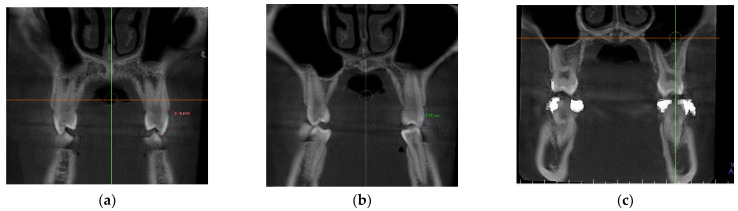
The purpose of this study was to evaluate skeletal, dentoalveolar and dental changes after Mini-screw Assisted Rapid Palatal Expansion (MARPE) using tooth bone-borne expanders in adolescent patients after analyzing different craniofacial references by Cone beam computed tomography (CBCT) and digital model analysis. This prospective, non-controlled intervention study was conducted on fifteen subjects (mean age 17 ± 4 years) with transversal maxillary deficiency. Pre (T1) and post-expansion (T2) CBCTs and casts were taken to evaluate changes at the premolars and first molar areas. To compare means between two times, paired samples t- or Wilcoxon test were used following criteria. Significant skeletal changes were found after treatment for Nasal width and Maxillary width with means of 2.1 (1.1) mm and 2.5 (1.6) mm (p < 0.00005). Midpalatal suture showed a tendency of parallel suture opening in the axial and coronal view. For dentoalveolar changes, a significant but small buccal bone thickness (BBT) reduction was observed in all teeth with a mean reduction of 0.3 mm for the right and left sides, especially for the distobuccal root of the first molar on the left side (DBBTL1M) [IC95%: (−0.6; −0.2); p = 0.001] with 0.4 (0.4) mm. However, a significant augmentation was observed for the palatal bone thickness (PBT) on the left side. The buccal alveolar crest (BACL) and dental inclination (DI) showed no significant changes after treatment in all the evaluated teeth. MARPE using tooth bone-borne appliances can achieve successful skeletal transverse maxillary expansion in adolescent patients, observing small dentoalveolar changes as buccal bone thickness (BBT) reduction, which was not clinically detectable. Most maxillary expansions derived from skeletal expansion, keeping the alveolar bone almost intact with minor buccal dental tipping.
本研究的目的是在通过锥形束计算机断层扫描(CBCT)和数字模型分析对不同颅面参考进行分析后,评估青少年患者使用牙骨支撑扩弓器进行微型螺钉辅助快速腭中缝扩展(MARPE)后的骨骼、牙槽骨和牙齿变化。本前瞻性、非对照干预研究针对15名横向上颌骨发育不足的受试者(平均年龄17±4岁)开展。在扩弓前(T1)和扩弓后(T2)拍摄CBCT和模型,以评估前磨牙和第一磨牙区域的变化。根据标准,使用配对样本t检验或Wilcoxon检验比较两次测量的均值。治疗后,鼻宽和上颌骨宽度出现显著的骨骼变化,均值分别为2.1(1.1)mm和2.5(1.6)mm(p<0.00005)。腭中缝在轴向和冠状位显示出平行缝打开的趋势。对于牙槽骨变化,所有牙齿的颊侧骨厚度(BBT)均有显著但微小的减少,左右两侧平均减少0.3mm,尤其是左侧第一磨牙远中颊根(DBBTL1M)[IC95%:(-0.6;-0.2);p=0.001]减少0.4(0.4)mm。然而,左侧腭侧骨厚度(PBT)出现显著增加。所有评估牙齿的颊侧牙槽嵴(BACL)和牙齿倾斜度(DI)在治疗后均无显著变化。使用牙骨支撑矫治器的MARPE可在青少年患者中成功实现上颌骨横向骨骼扩展,观察到牙槽骨变化较小,如颊侧骨厚度(BBT)减少,这在临床上无法检测到。大多数上颌骨扩展源于骨骼扩展,牙槽骨几乎保持完整,牙齿仅有轻微的颊侧倾斜。