Department of Pathology, Forensic Medicine and Cytology, University Hospital Centre Split, 21000 Split, Croatia.
Department of Anatomy, Histology and Embryology, School of Medicine, University of Split, 21000 Split, Croatia.
Medicina (Kaunas). 2022 Aug 21;58(8):1135. doi: 10.3390/medicina58081135.
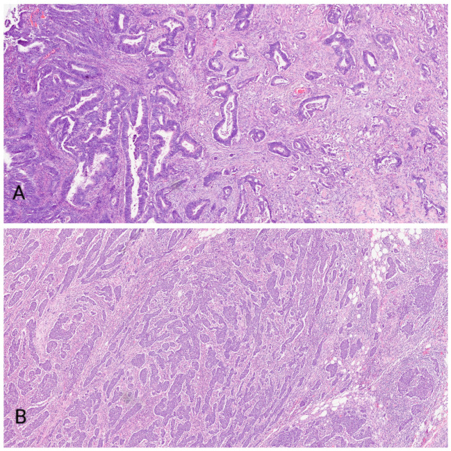
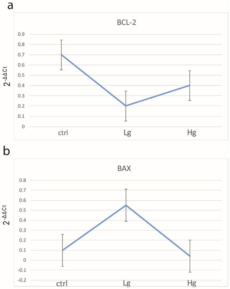
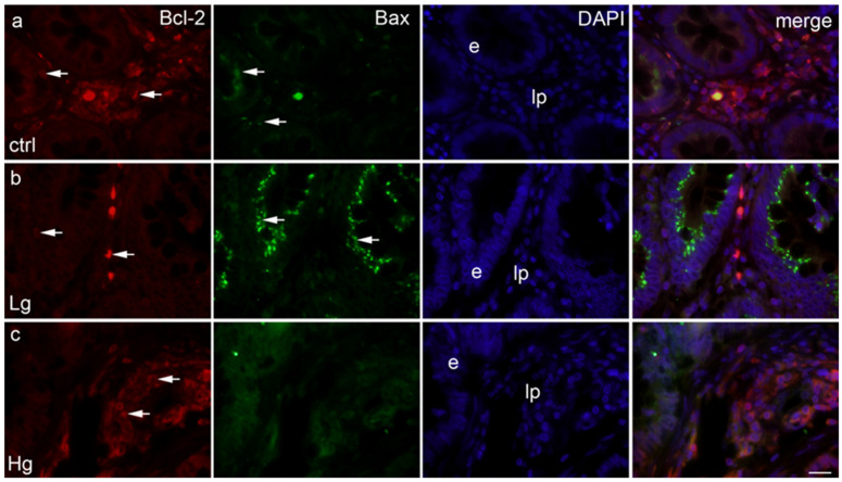
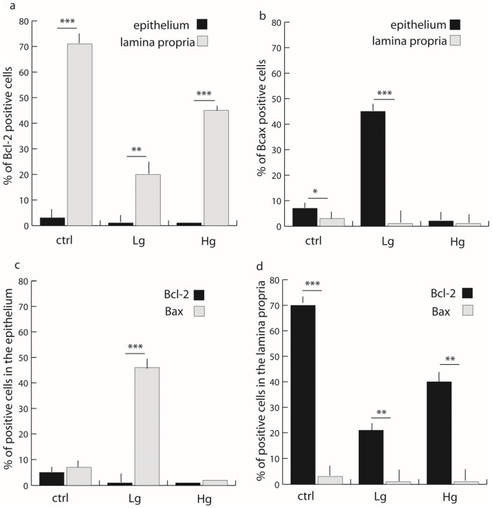
The epithelial and stromal tissues both play a role in the progression of colorectal cancer (CRC). The aim of this study was to assess the expression of anti-apoptotic Bcl-2 and pro-apoptotic Bax in the epithelium as well as the lamina propria of normal colonic controls, low-grade tumor samples and high-grade tumor samples. A total of 60 samples consisting of both normal colonic and carcinoma samples was collected from the Department of Pathology, Cytology and Forensic Medicine, University Hospital Center, Split from January 2020 to December 2021. The expression of Bcl-2 and Bax markers was semi-quantitatively and quantitatively evaluated by recording immunofluorescence stain intensity and by counting stained cells in the lamina propria and epithelium. Analysis of positive cells was performed using the Mann-Whitney test. In all samples, Bcl-2 was significantly more expressed in the lamina propria when compared with the epithelium. Bax was significantly more expressed in the epithelium of normal and low-grade cancer samples when compared with their respective laminae propriae. The percentage of Bcl-2-positive cells in lamina propria is about two times lower in high-grade CRC and about three times lower in low-grade CRC in comparison with healthy controls. Contrary to this, the percentage of Bax-positive cells was greater in the epithelium of low-grade CRC in comparison with healthy control and high-grade CRC. Our study provides a new insight into Bcl-2 and Bax expression pattern in CRC. Evaluation of Bcl-2 expression in the lamina propria and Bax expression in the epithelium could provide important information for colorectal cancer prognosis as well as potential treatment strategies.
上皮组织和基质组织在结直肠癌(CRC)的进展中都发挥了作用。本研究旨在评估抗凋亡 Bcl-2 和促凋亡 Bax 在正常结肠对照、低级别肿瘤样本和高级别肿瘤样本的上皮和固有层中的表达。
2020 年 1 月至 2021 年 12 月,从斯普利特大学医院中心病理学、细胞学和法医学系收集了 60 个包含正常结肠和癌样本的样本。通过记录免疫荧光染色强度并对固有层和上皮中的染色细胞进行计数,对 Bcl-2 和 Bax 标志物的表达进行半定量和定量评估。使用曼-惠特尼检验对阳性细胞进行分析。
在所有样本中,Bcl-2 在固有层中的表达明显高于上皮。与各自的固有层相比,Bax 在正常和低级别癌样本的上皮中的表达明显更高。与健康对照组相比,高级别 CRC 固有层中 Bcl-2 阳性细胞的百分比约低两倍,低级别 CRC 固有层中 Bcl-2 阳性细胞的百分比约低三倍。与此相反,与健康对照组和高级别 CRC 相比,低级别 CRC 上皮中的 Bax 阳性细胞百分比更大。
我们的研究为 CRC 中 Bcl-2 和 Bax 表达模式提供了新的见解。固有层中 Bcl-2 的表达和上皮中 Bax 的表达评估可为结直肠癌的预后以及潜在的治疗策略提供重要信息。