Department of Biochemistry and Pharmacology, Bio21 Molecular Science and Biotechnology Institute, The University of Melbourne, Parkville, VIC, 3010, Australia.
Biological Optical Microscopy Platform, The University of Melbourne, Parkville, VIC, 3010, Australia.
Nat Commun. 2022 Aug 27;13(1):5054. doi: 10.1038/s41467-022-32579-4.
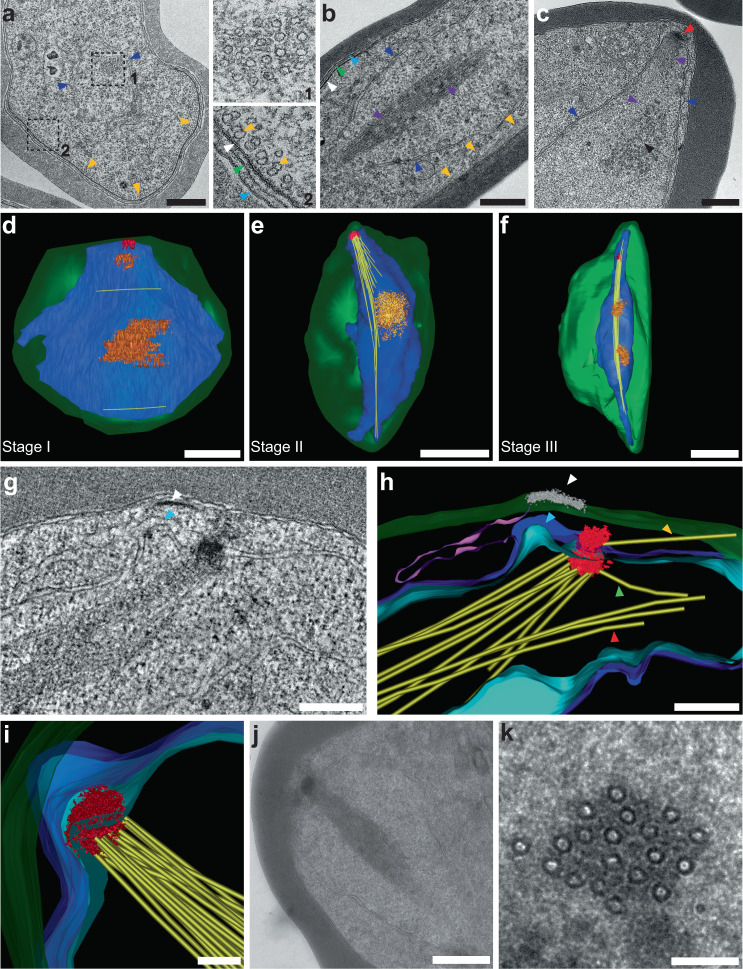
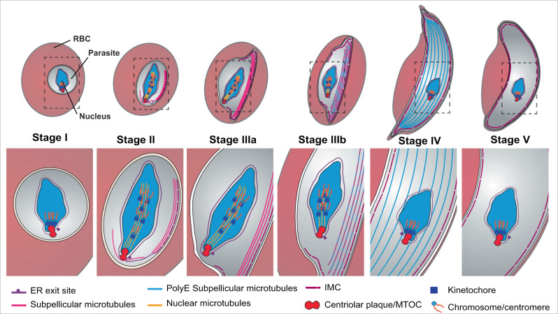
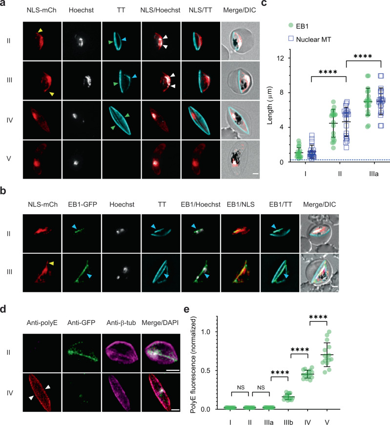
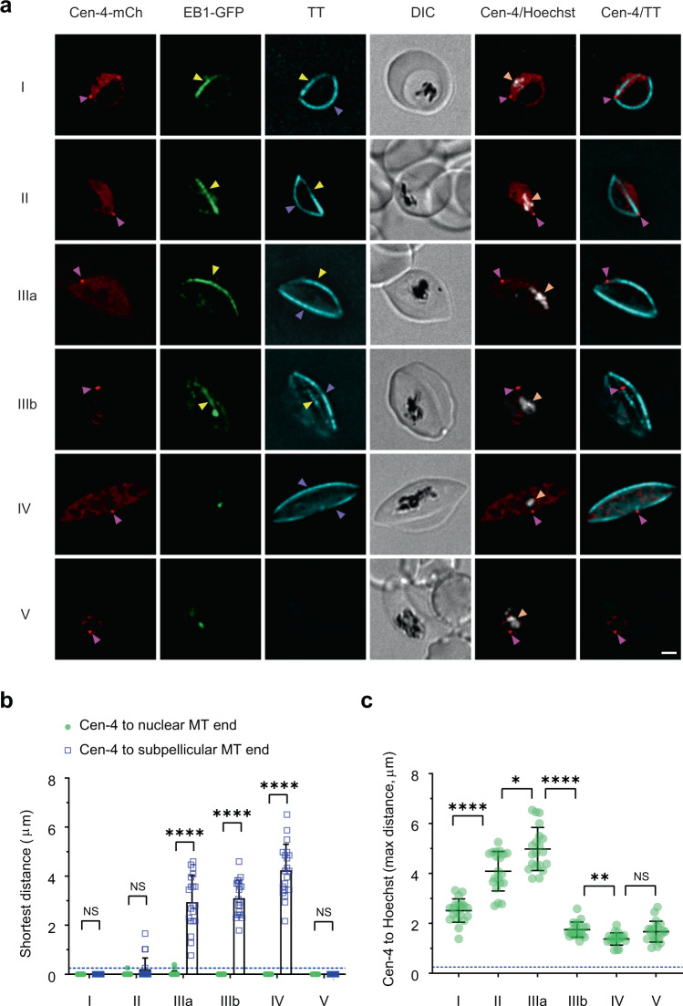
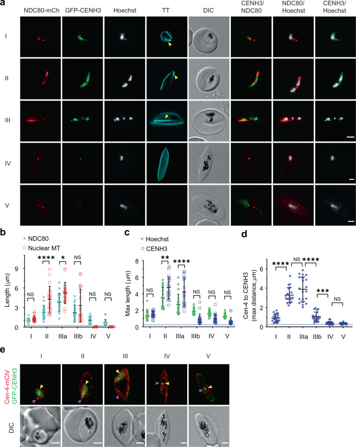
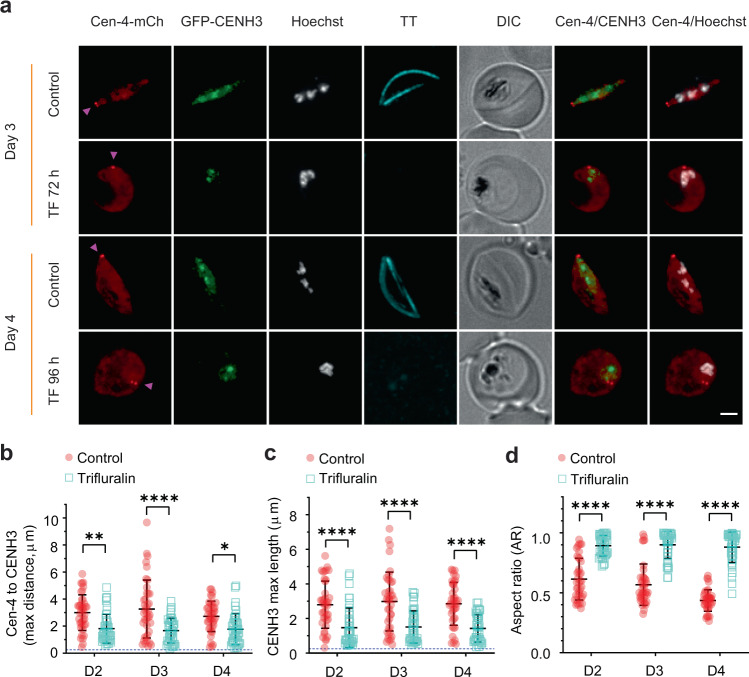
The sexual stage gametocytes of the malaria parasite, Plasmodium falciparum, adopt a falciform (crescent) shape driven by the assembly of a network of microtubules anchored to a cisternal inner membrane complex (IMC). Using 3D electron microscopy, we show that a non-mitotic microtubule organizing center (MTOC), embedded in the parasite's nuclear membrane, orients the endoplasmic reticulum and the nascent IMC and seeds cytoplasmic microtubules. A bundle of microtubules extends into the nuclear lumen, elongating the nuclear envelope and capturing the chromatin. Classical mitotic machinery components, including centriolar plaque proteins, Pfcentrin-1 and -4, microtubule-associated protein, End-binding protein-1, kinetochore protein, PfNDC80 and centromere-associated protein, PfCENH3, are involved in the nuclear microtubule assembly/disassembly process. Depolymerisation of the microtubules using trifluralin prevents elongation and disrupts the chromatin, centromere and kinetochore organisation. We show that the unusual non-mitotic hemispindle plays a central role in chromatin organisation, IMC positioning and subpellicular microtubule formation in gametocytes.
疟原虫(Plasmodium falciparum)的有性生殖阶段配子体,通过锚定在质膜内层复合物(IMC)上的微管网络的组装,采用镰刀形(新月形)形状。使用 3D 电子显微镜,我们表明,嵌入寄生虫核膜中的非有丝分裂微管组织中心(MTOC),使内质网和新生 IMC 以及细胞质微管定向。一束微管延伸到核腔中,使核膜拉长并捕获染色质。经典的有丝分裂机制成分,包括中心粒斑蛋白 Pfcentrin-1 和 -4、微管相关蛋白 End-binding protein-1、动粒蛋白 PfNDC80 和着丝粒相关蛋白 PfCENH3,参与核微管组装/解组装过程。使用三氟拉林使微管解聚会阻止伸长并破坏染色质、着丝粒和动粒的组织。我们表明,这种不寻常的非有丝分裂半纺锤体在配子体的染色质组织、IMC 定位和皮层下微管形成中起着核心作用。