• 文献检索
  • 文档翻译
  • 深度研究
  • 学术资讯
  • Suppr Zotero 插件Zotero 插件
  • 邀请有礼
  • 套餐&价格
  • 历史记录
应用&插件
Suppr Zotero 插件Zotero 插件浏览器插件Mac 客户端Windows 客户端微信小程序
定价
高级版会员购买积分包购买API积分包
服务
文献检索文档翻译深度研究API 文档MCP 服务
关于我们
关于 Suppr公司介绍联系我们用户协议隐私条款
关注我们

Suppr 超能文献

核心技术专利:CN118964589B侵权必究
粤ICP备2023148730 号-1Suppr @ 2026

文献检索

告别复杂PubMed语法,用中文像聊天一样搜索,搜遍4000万医学文献。AI智能推荐,让科研检索更轻松。

立即免费搜索

文件翻译

保留排版,准确专业,支持PDF/Word/PPT等文件格式,支持 12+语言互译。

免费翻译文档

深度研究

AI帮你快速写综述,25分钟生成高质量综述,智能提取关键信息,辅助科研写作。

立即免费体验

基于累积顺序保持图像变换的变分自动编码器在眼前节光学相干断层扫描图像中的应用。

Development of Cumulative Order-Preserving Image Transformation Based Variational Autoencoder for Anterior Segment Optical Coherence Tomography Images.

机构信息

Department of Ophthalmology, Gangneung Asan Hospital, Gangneung, Korea.

Asan Artificial Intelligence Institute, Hwaseong-si, Gyeonggi-do, Korea.

出版信息

Transl Vis Sci Technol. 2022 Aug 1;11(8):30. doi: 10.1167/tvst.11.8.30.

DOI:10.1167/tvst.11.8.30
PMID:36040250
原文链接:https://pmc.ncbi.nlm.nih.gov/articles/PMC9437650/
Abstract

PURPOSE

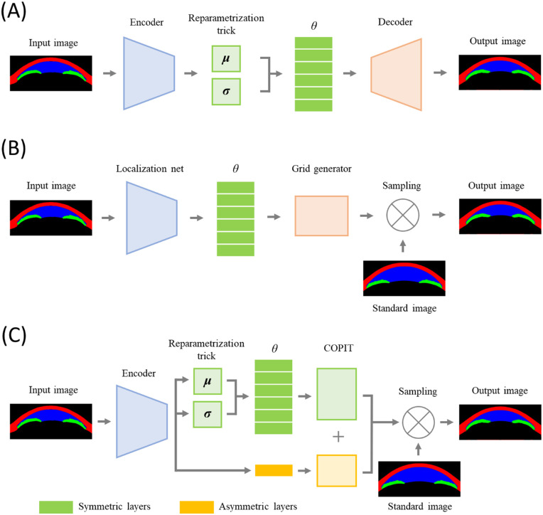
To develop a variational autoencoder (VAE) suitable for analysis of the latent structure of anterior segment optical coherence tomography (AS-OCT) images and to investigate possibilities of latent structure analysis of the AS-OCT images.

METHODS

We retrospectively collected clinical data and AS-OCT images from 2111 eyes of 1261 participants from the ongoing Asan Glaucoma Progression Study. A specifically modified VAE was used to extract six symmetrical and one asymmetrical latent variable. A total of 1692 eyes of 1007 patients were used for training the model. Conventional measurements and latent variables were compared between 74 primary angle closure (PAC) and 51 primary angle closure glaucoma (PACG) eyes from validation set (419 eyes of 254 patients) that were not used for training.

RESULTS

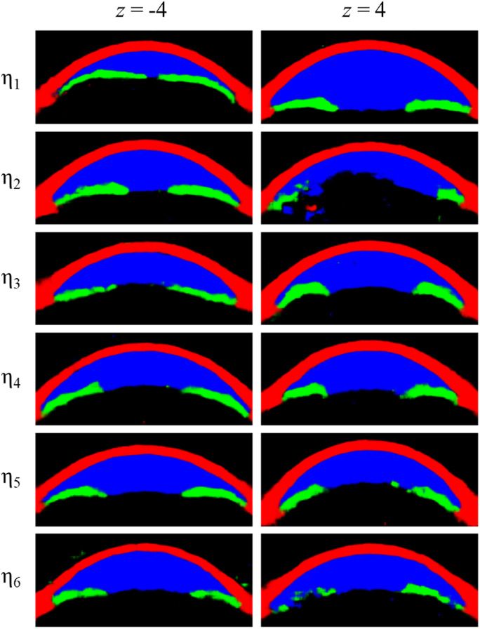
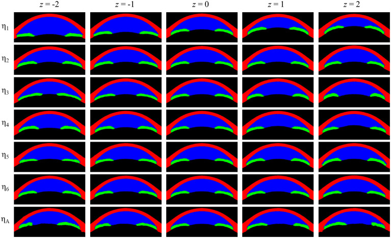
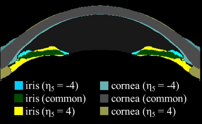
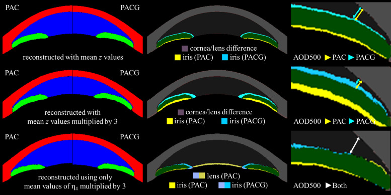
Among the symmetrical latent variables, the first three and the last demonstrated easily recognized features, anterior chamber area in η1, curvature of the cornea in η2, the pupil size in η3 and corneal thickness in η6, whereas η4 and η5 were more complex aggregating complex interactions of multiple structures. Compared with PAC eyes, there was no difference in any of the conventional measurements in PACG eyes. However, values of η4 were significantly different between the two groups, being smaller in the PACG group (P = 0.015).

CONCLUSIONS

VAE is a useful framework for analysis of the latent structure of AS-OCT. Latent structure analysis could be useful in capturing features not readily evident with conventional measures.

TRANSLATIONAL RELEVANCE

This study suggested that a deep learning-based latent space model can be applied for the analysis of AS-OCT images to find latent characteristics of the anterior segment of the eye.

摘要

目的

开发一种变分自动编码器(VAE),适用于分析眼前节光学相干断层扫描(AS-OCT)图像的潜在结构,并探讨对 AS-OCT 图像进行潜在结构分析的可能性。

方法

我们回顾性地收集了正在进行的 Asan 青光眼进展研究中 1261 名参与者的 2111 只眼的临床数据和 AS-OCT 图像。使用专门修改的 VAE 提取 6 个对称和 1 个非对称的潜在变量。共使用 1007 名患者的 1692 只眼对模型进行训练。在未用于训练的验证集(419 只眼,254 名患者)中,将 74 只原发性闭角型青光眼(PAC)和 51 只原发性闭角型青光眼(PACG)眼的常规测量值和潜在变量与 51 只原发性闭角型青光眼(PACG)眼进行比较。

结果

在对称的潜在变量中,前三个和最后一个变量具有易于识别的特征,η1 中的前房面积、η2 中的角膜曲率、η3 中的瞳孔大小和η6 中的角膜厚度,而η4 和η5 则更复杂,综合了多个结构的复杂相互作用。与 PAC 眼相比,PACG 眼中的任何常规测量值均无差异。然而,两组间η4 的值差异有统计学意义,PACG 组较小(P=0.015)。

结论

VAE 是分析 AS-OCT 潜在结构的有用框架。潜在结构分析可能有助于捕捉用常规测量方法不易发现的特征。

翻译的准确性和流畅性都比较高,能够准确表达原文的意思。

https://cdn.ncbi.nlm.nih.gov/pmc/blobs/9433/9437650/dc61ed0f8ce7/tvst-11-8-30-f005.jpg
https://cdn.ncbi.nlm.nih.gov/pmc/blobs/9433/9437650/d68825c5dae9/tvst-11-8-30-f001.jpg
https://cdn.ncbi.nlm.nih.gov/pmc/blobs/9433/9437650/89288637861c/tvst-11-8-30-f002.jpg
https://cdn.ncbi.nlm.nih.gov/pmc/blobs/9433/9437650/9238c42e0c65/tvst-11-8-30-f003.jpg
https://cdn.ncbi.nlm.nih.gov/pmc/blobs/9433/9437650/b1d0668e0b70/tvst-11-8-30-f004.jpg
https://cdn.ncbi.nlm.nih.gov/pmc/blobs/9433/9437650/dc61ed0f8ce7/tvst-11-8-30-f005.jpg

相似文献

1
Development of Cumulative Order-Preserving Image Transformation Based Variational Autoencoder for Anterior Segment Optical Coherence Tomography Images.基于累积顺序保持图像变换的变分自动编码器在眼前节光学相干断层扫描图像中的应用。
Transl Vis Sci Technol. 2022 Aug 1;11(8):30. doi: 10.1167/tvst.11.8.30.
2
Development of a β-Variational Autoencoder for Disentangled Latent Space Representation of Anterior Segment Optical Coherence Tomography Images.用于前节光学相干断层扫描图像去纠缠潜在空间表示的 β 变分自动编码器的开发。
Transl Vis Sci Technol. 2022 Feb 1;11(2):11. doi: 10.1167/tvst.11.2.11.
3
Deep Learning-Based Classification of Subtypes of Primary Angle-Closure Disease With Anterior Segment Optical Coherence Tomography.基于深度学习的前节段光学相干断层扫描对原发性闭角型青光眼亚型的分类。
J Glaucoma. 2023 Jun 1;32(6):540-547. doi: 10.1097/IJG.0000000000002194. Epub 2023 Mar 3.
4
[Detection of the anterior segment configuration of primary angle-closure glaucoma by anterior segment optical coherence tomography].[眼前节光学相干断层扫描对原发性闭角型青光眼眼前节形态的检测]
Zhonghua Yan Ke Za Zhi. 2013 Feb;49(2):109-15.
5
Evaluation of the Diagnostic Performance of Swept-Source Anterior Segment Optical Coherence Tomography in Primary Angle Closure Disease.原发性闭角型青光眼的扫频源眼前节光学相干断层扫描诊断性能评估。
Am J Ophthalmol. 2022 Jan;233:68-77. doi: 10.1016/j.ajo.2021.06.033. Epub 2021 Jul 17.
6
The anterior and posterior biometric characteristics in primary angle-closure disease: Data based on anterior segment optical coherence tomography and swept-source optical coherence tomography.原发性闭角型青光眼的前房和后房生物测量特征:基于眼前节光学相干断层扫描和扫频源光学相干断层扫描的数据。
Indian J Ophthalmol. 2021 Apr;69(4):865-870. doi: 10.4103/ijo.IJO_936_20.
7
A hierarchical cluster analysis of primary angle closure classification using anterior segment optical coherence tomography parameters.应用眼前节光学相干断层扫描参数对原发性闭角型青光眼分类的层次聚类分析。
Invest Ophthalmol Vis Sci. 2013 Jan 30;54(1):848-53. doi: 10.1167/iovs.12-10391.
8
Limbus-insertion distance of superior rectus in primary angle closure glaucoma: an anterior segment OCT study.原发性闭角型青光眼患者上直肌角膜缘插入距离:一项眼前节光学相干断层扫描研究
Can J Ophthalmol. 2016 Dec;51(6):438-444. doi: 10.1016/j.jcjo.2016.06.003. Epub 2016 Oct 1.
9
Outcomes of laser peripheral iridotomy in angle closure subgroups according to anterior segment optical coherence tomography parameters.根据眼前节光学相干断层扫描参数,激光周边虹膜切开术在闭角型亚组中的治疗效果。
Invest Ophthalmol Vis Sci. 2014 Sep 23;55(10):6795-801. doi: 10.1167/iovs.14-14714.
10
Detection of primary angle closure using anterior segment optical coherence tomography in Asian eyes.在亚洲人眼中使用眼前节光学相干断层扫描检测原发性闭角型青光眼
Ophthalmology. 2007 Jan;114(1):33-9. doi: 10.1016/j.ophtha.2006.05.073. Epub 2006 Oct 27.

引用本文的文献

1
Automated spectral decomposition and reconstruction of optical properties using a mixed autoencoder approach.使用混合自动编码器方法对光学特性进行自动光谱分解和重建。
J Biomed Opt. 2025 Apr;30(4):047001. doi: 10.1117/1.JBO.30.4.047001. Epub 2025 Apr 9.
2
Quantifying the spatial patterns of retinal ganglion cell loss and progression in optic neuropathy by applying a deep learning variational autoencoder approach to optical coherence tomography.通过将深度学习变分自编码器方法应用于光学相干断层扫描来量化视神经病变中视网膜神经节细胞丢失和进展的空间模式。
Front Ophthalmol (Lausanne). 2025 Feb 3;4:1497848. doi: 10.3389/fopht.2024.1497848. eCollection 2024.
3

本文引用的文献

1
Development of a β-Variational Autoencoder for Disentangled Latent Space Representation of Anterior Segment Optical Coherence Tomography Images.用于前节光学相干断层扫描图像去纠缠潜在空间表示的 β 变分自动编码器的开发。
Transl Vis Sci Technol. 2022 Feb 1;11(2):11. doi: 10.1167/tvst.11.2.11.
2
Angle-closure assessment in anterior segment OCT images via deep learning.基于深度学习的眼前节 OCT 图像的闭角评估。
Med Image Anal. 2021 Apr;69:101956. doi: 10.1016/j.media.2021.101956. Epub 2021 Jan 7.
3
Deep Learning for Anterior Segment Optical Coherence Tomography to Predict the Presence of Plateau Iris.
Artificial Intelligence for Optical Coherence Tomography in Glaucoma.
用于青光眼光学相干断层扫描的人工智能
Transl Vis Sci Technol. 2025 Jan 2;14(1):27. doi: 10.1167/tvst.14.1.27.
4
The application of artificial intelligence in glaucoma diagnosis and prediction.人工智能在青光眼诊断与预测中的应用。
Front Cell Dev Biol. 2023 May 4;11:1173094. doi: 10.3389/fcell.2023.1173094. eCollection 2023.
用于眼前节光学相干断层扫描的深度学习以预测高原虹膜的存在
Transl Vis Sci Technol. 2021 Jan 6;10(1):7. doi: 10.1167/tvst.10.1.7. eCollection 2021 Jan.
4
The Use of Anterior-Segment Optical-Coherence Tomography for the Assessment of the Iridocorneal Angle and Its Alterations: Update and Current Evidence.眼前节光学相干断层扫描在虹膜角膜角评估及其改变中的应用:更新与当前证据
J Clin Med. 2021 Jan 11;10(2):231. doi: 10.3390/jcm10020231.
5
Deep learning algorithms to isolate and quantify the structures of the anterior segment in optical coherence tomography images.深度学习算法在光学相干断层扫描图像中分离和量化前段结构。
Br J Ophthalmol. 2021 Sep;105(9):1231-1237. doi: 10.1136/bjophthalmol-2019-315723. Epub 2020 Sep 26.
6
Deep Neural Network for Scleral Spur Detection in Anterior Segment OCT Images: The Chinese American Eye Study.深层神经网络在眼前节 OCT 图像中巩膜突检测的应用:中美眼研究。
Transl Vis Sci Technol. 2020 Mar 30;9(2):18. doi: 10.1167/tvst.9.2.18. eCollection 2020 Mar.
7
A Deep Learning System for Automated Angle-Closure Detection in Anterior Segment Optical Coherence Tomography Images.基于深度学习的眼前节光学相干断层扫描图像中房角关闭自动检测系统。
Am J Ophthalmol. 2019 Jul;203:37-45. doi: 10.1016/j.ajo.2019.02.028. Epub 2019 Mar 6.
8
Classification of primary angle closure spectrum with hierarchical cluster analysis.基于层次聚类分析的原发性闭角型青光眼分类。
PLoS One. 2018 Jul 23;13(7):e0199157. doi: 10.1371/journal.pone.0199157. eCollection 2018.
9
Long-term Changes in Anterior Segment Characteristics of Eyes With Different Primary Angle-Closure Mechanisms.不同原发性闭角型青光眼机制眼球前段特征的长期变化。
Am J Ophthalmol. 2018 Jul;191:54-63. doi: 10.1016/j.ajo.2018.04.005. Epub 2018 Apr 12.
10
Subclassification of Primary Angle Closure Using Anterior Segment Optical Coherence Tomography and Ultrasound Biomicroscopic Parameters.使用眼前节光学相干断层扫描和超声生物显微镜参数对原发性闭角型青光眼进行亚分类。
Ophthalmology. 2017 Jul;124(7):1039-1047. doi: 10.1016/j.ophtha.2017.02.025. Epub 2017 Apr 3.