• 文献检索
  • 文档翻译
  • 深度研究
  • 学术资讯
  • Suppr Zotero 插件Zotero 插件
  • 邀请有礼
  • 套餐&价格
  • 历史记录
应用&插件
Suppr Zotero 插件Zotero 插件浏览器插件Mac 客户端Windows 客户端微信小程序
定价
高级版会员购买积分包购买API积分包
服务
文献检索文档翻译深度研究API 文档MCP 服务
关于我们
关于 Suppr公司介绍联系我们用户协议隐私条款
关注我们

Suppr 超能文献

核心技术专利:CN118964589B侵权必究
粤ICP备2023148730 号-1Suppr @ 2026

文献检索

告别复杂PubMed语法,用中文像聊天一样搜索,搜遍4000万医学文献。AI智能推荐,让科研检索更轻松。

立即免费搜索

文件翻译

保留排版,准确专业,支持PDF/Word/PPT等文件格式,支持 12+语言互译。

免费翻译文档

深度研究

AI帮你快速写综述,25分钟生成高质量综述,智能提取关键信息,辅助科研写作。

立即免费体验

基于机器学习的脑 CT 图像皮质萎缩自动评估。

Machine learning-based automatic estimation of cortical atrophy using brain computed tomography images.

机构信息

Department of Neurology, School of Medicine, Kangwon National University, Chuncheon, Republic of Korea.

Department of Neurology, Kangwon National University Hospital, Chuncheon, Republic of Korea.

出版信息

Sci Rep. 2022 Aug 30;12(1):14740. doi: 10.1038/s41598-022-18696-6.

DOI:10.1038/s41598-022-18696-6
PMID:36042322
原文链接:https://pmc.ncbi.nlm.nih.gov/articles/PMC9427760/
Abstract

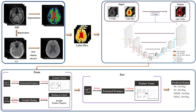
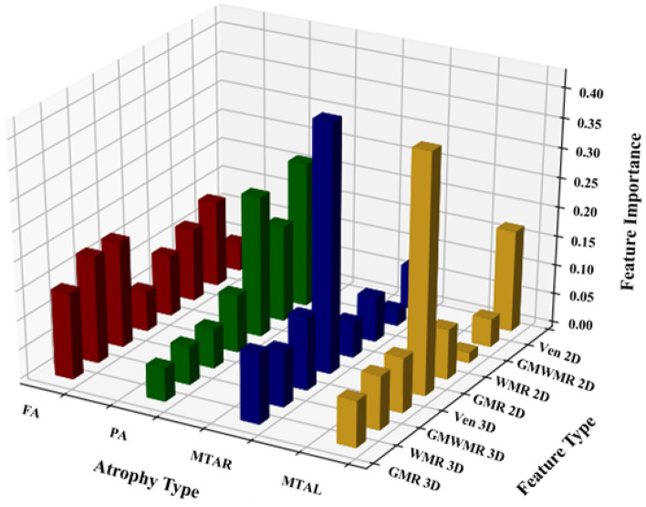
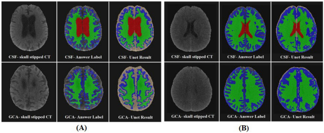
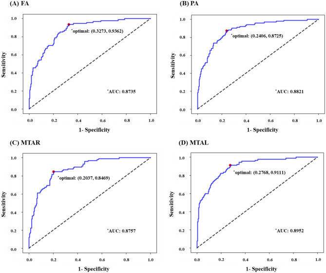
Cortical atrophy is measured clinically according to established visual rating scales based on magnetic resonance imaging (MRI). Although brain MRI is the primary imaging marker for neurodegeneration, computed tomography (CT) is also widely used for the early detection and diagnosis of dementia. However, they are seldom investigated. Therefore, we developed a machine learning algorithm for the automatic estimation of cortical atrophy on brain CT. Brain CT images (259 Alzheimer's dementia and 55 cognitively normal subjects) were visually rated by three neurologists and used for training. We constructed an algorithm by combining the convolutional neural network and regularized logistic regression (RLR). Model performance was then compared with that of neurologists, and feature importance was measured. RLR provided fast and reliable automatic estimations of frontal atrophy (75.2% accuracy, 93.6% sensitivity, 67.2% specificity, and 0.87 area under the curve [AUC]), posterior atrophy (79.6% accuracy, 87.2% sensitivity, 75.9% specificity, and 0.88 AUC), right medial temporal atrophy (81.2% accuracy, 84.7% sensitivity, 79.6% specificity, and 0.88 AUC), and left medial temporal atrophy (77.7% accuracy, 91.1% sensitivity, 72.3% specificity, and 0.90 AUC). We concluded that RLR-based automatic estimation of brain CT provided a comprehensive rating of atrophy that can potentially support physicians in real clinical settings.

摘要

皮质萎缩是根据磁共振成像(MRI)的既定视觉评估量表在临床上进行测量的。虽然脑 MRI 是神经退行性变的主要影像学标志物,但计算机断层扫描(CT)也广泛用于痴呆的早期检测和诊断。然而,它们很少被研究。因此,我们开发了一种用于自动估计脑 CT 皮质萎缩的机器学习算法。脑 CT 图像(259 例阿尔茨海默病痴呆和 55 例认知正常受试者)由三位神经科医生进行视觉评估,并用于训练。我们通过结合卷积神经网络和正则化逻辑回归(RLR)构建了一个算法。然后将模型性能与神经科医生进行比较,并测量特征重要性。RLR 可快速可靠地自动估计额部萎缩(准确率 75.2%,敏感性 93.6%,特异性 67.2%,曲线下面积 [AUC] 为 0.87)、后部萎缩(准确率 79.6%,敏感性 87.2%,特异性 75.9%,AUC 为 0.88)、右侧内侧颞叶萎缩(准确率 81.2%,敏感性 84.7%,特异性 79.6%,AUC 为 0.88)和左侧内侧颞叶萎缩(准确率 77.7%,敏感性 91.1%,特异性 72.3%,AUC 为 0.90)。我们得出结论,基于 RLR 的脑 CT 自动估计可提供全面的萎缩评分,可能有助于医生在实际临床环境中做出决策。

https://cdn.ncbi.nlm.nih.gov/pmc/blobs/0102/9427760/beef8c4dd3e1/41598_2022_18696_Fig4_HTML.jpg
https://cdn.ncbi.nlm.nih.gov/pmc/blobs/0102/9427760/0b02290dfbc6/41598_2022_18696_Fig1_HTML.jpg
https://cdn.ncbi.nlm.nih.gov/pmc/blobs/0102/9427760/6677dd696bc5/41598_2022_18696_Fig2_HTML.jpg
https://cdn.ncbi.nlm.nih.gov/pmc/blobs/0102/9427760/734fd363fb69/41598_2022_18696_Fig3_HTML.jpg
https://cdn.ncbi.nlm.nih.gov/pmc/blobs/0102/9427760/beef8c4dd3e1/41598_2022_18696_Fig4_HTML.jpg

相似文献

1
Machine learning-based automatic estimation of cortical atrophy using brain computed tomography images.基于机器学习的脑 CT 图像皮质萎缩自动评估。
Sci Rep. 2022 Aug 30;12(1):14740. doi: 10.1038/s41598-022-18696-6.
2
AVRA: Automatic visual ratings of atrophy from MRI images using recurrent convolutional neural networks.AVRA:基于递归卷积神经网络的 MRI 图像自动萎缩评分。
Neuroimage Clin. 2019;23:101872. doi: 10.1016/j.nicl.2019.101872. Epub 2019 May 25.
3
Structural magnetic resonance imaging for the early diagnosis of dementia due to Alzheimer's disease in people with mild cognitive impairment.用于轻度认知障碍患者阿尔茨海默病所致痴呆早期诊断的结构磁共振成像
Cochrane Database Syst Rev. 2020 Mar 2;3(3):CD009628. doi: 10.1002/14651858.CD009628.pub2.
4
Determination of Alzheimer's disease based on morphology and atrophy using machine learning combined with automated segmentation.基于机器学习结合自动分割的形态学和萎缩分析诊断阿尔茨海默病。
Acta Radiol. 2024 Apr;65(4):359-366. doi: 10.1177/02841851231218384. Epub 2024 Jan 9.
5
Regional MRI volumetry using NeuroQuant versus visual rating scales in patients with cognitive impairment and dementia.使用 NeuroQuant 进行区域性 MRI 容积测量与认知障碍和痴呆患者的视觉评分量表相比。
Brain Behav. 2024 Feb;14(2):e3397. doi: 10.1002/brb3.3397.
6
Accurate prediction of histologically confirmed Alzheimer's disease and the differential diagnosis of dementia: the use of NINCDS-ADRDA and DSM-III-R criteria, SPECT, X-ray CT, and APO E4 medial temporal lobe dementias. The Oxford Project to Investigate Memory and Aging.组织学确诊的阿尔茨海默病的准确预测及痴呆的鉴别诊断:采用NINCDS - ADRDA和DSM - III - R标准、单光子发射计算机断层扫描(SPECT)、X线计算机断层扫描(X-ray CT)以及载脂蛋白E4(APO E4)与内侧颞叶痴呆。牛津记忆与衰老研究项目。
Int Psychogeriatr. 1997;9 Suppl 1:191-222; discussion 247-52.
7
A new age-related cutoff of medial temporal atrophy scale on MRI improving the diagnostic accuracy of neurodegeneration due to Alzheimer's disease in a Chinese population.MRI 上与年龄相关的内侧颞叶萎缩量表新界值可提高中国人群阿尔茨海默病所致神经退行性变的诊断准确性。
BMC Geriatr. 2019 Feb 28;19(1):59. doi: 10.1186/s12877-019-1072-8.
8
A novel CT-based automated analysis method provides comparable results with MRI in measuring brain atrophy and white matter lesions.一种新的基于 CT 的自动分析方法在测量脑萎缩和白质病变方面提供了与 MRI 可比的结果。
Neuroradiology. 2021 Dec;63(12):2035-2046. doi: 10.1007/s00234-021-02761-4. Epub 2021 Aug 14.
9
Brain-on-Cloud for automatic diagnosis of Alzheimer's disease from 3D structural magnetic resonance whole-brain scans.基于云的脑成像技术用于从 3D 结构磁共振全脑扫描中自动诊断阿尔茨海默病。
Comput Methods Programs Biomed. 2022 Dec;227:107191. doi: 10.1016/j.cmpb.2022.107191. Epub 2022 Oct 27.
10
Selecting the most relevant brain regions to discriminate Alzheimer's disease patients from healthy controls using multiple kernel learning: A comparison across functional and structural imaging modalities and atlases.使用多核学习选择最相关的大脑区域来区分阿尔茨海默病患者与健康对照者:功能和结构成像方式及图谱的比较。
Neuroimage Clin. 2017 Nov 9;17:628-641. doi: 10.1016/j.nicl.2017.10.026. eCollection 2018.

引用本文的文献

1
The diagnostic accuracy of CTseg segmentation software for dementia in a New Zealand memory service.CTseg分割软件在新西兰记忆服务中对痴呆症的诊断准确性。
J Alzheimers Dis Rep. 2025 May 21;9:25424823251332448. doi: 10.1177/25424823251332448. eCollection 2025 Jan-Dec.
2
Neuroimaging techniques, gene therapy, and gut microbiota: frontier advances and integrated applications in Alzheimer's Disease research.神经成像技术、基因治疗与肠道微生物群:阿尔茨海默病研究中的前沿进展与综合应用
Front Aging Neurosci. 2024 Dec 3;16:1485657. doi: 10.3389/fnagi.2024.1485657. eCollection 2024.

本文引用的文献

1
Development and Validation of a Deep Learning-Based Automatic Brain Segmentation and Classification Algorithm for Alzheimer Disease Using 3D T1-Weighted Volumetric Images.基于深度学习的 3D T1 加权容积图像阿尔茨海默病自动脑分割与分类算法的建立与验证。
AJNR Am J Neuroradiol. 2020 Dec;41(12):2227-2234. doi: 10.3174/ajnr.A6848. Epub 2020 Nov 5.
2
On brain atlas choice and automatic segmentation methods: a comparison of MAPER & FreeSurfer using three atlas databases.脑图谱选择和自动分割方法:使用三个图谱数据库对 MAPER 和 FreeSurfer 的比较。
Sci Rep. 2020 Feb 18;10(1):2837. doi: 10.1038/s41598-020-57951-6.
3
Deep Learning in Alzheimer's Disease: Diagnostic Classification and Prognostic Prediction Using Neuroimaging Data.
阿尔茨海默病中的深度学习:利用神经影像数据进行诊断分类和预后预测
Front Aging Neurosci. 2019 Aug 20;11:220. doi: 10.3389/fnagi.2019.00220. eCollection 2019.
4
Clinical significance of amyloid β positivity in patients with probable cerebral amyloid angiopathy markers.淀粉样β阳性在可能有脑淀粉样血管病标志物的患者中的临床意义。
Eur J Nucl Med Mol Imaging. 2019 Jun;46(6):1287-1298. doi: 10.1007/s00259-019-04314-7. Epub 2019 Apr 2.
5
Machine learning of neuroimaging for assisted diagnosis of cognitive impairment and dementia: A systematic review.用于认知障碍和痴呆辅助诊断的神经影像机器学习:一项系统综述。
Alzheimers Dement (Amst). 2018 Aug 11;10:519-535. doi: 10.1016/j.dadm.2018.07.004. eCollection 2018.
6
A Nomogram for Predicting Amyloid PET Positivity in Amnestic Mild Cognitive Impairment.用于预测遗忘型轻度认知障碍淀粉样 PET 阳性的列线图。
J Alzheimers Dis. 2018;66(2):681-691. doi: 10.3233/JAD-180048.
7
NIA-AA Research Framework: Toward a biological definition of Alzheimer's disease.NIA-AA 研究框架:迈向阿尔茨海默病的生物学定义。
Alzheimers Dement. 2018 Apr;14(4):535-562. doi: 10.1016/j.jalz.2018.02.018.
8
A review on neuroimaging-based classification studies and associated feature extraction methods for Alzheimer's disease and its prodromal stages.基于神经影像学的阿尔茨海默病及其前驱期分类研究及相关特征提取方法综述。
Neuroimage. 2017 Jul 15;155:530-548. doi: 10.1016/j.neuroimage.2017.03.057. Epub 2017 Apr 13.
9
Comparison of automated brain segmentation using a brain phantom and patients with early Alzheimer's dementia or mild cognitive impairment.利用脑模型与早期阿尔茨海默病或轻度认知障碍患者对比自动脑分割。
Psychiatry Res. 2015 Sep 30;233(3):299-305. doi: 10.1016/j.pscychresns.2015.07.011. Epub 2015 Jul 17.
10
Automated CT-based segmentation and quantification of total intracranial volume.基于 CT 的全自动颅内总体积分割与定量。
Eur Radiol. 2015 Nov;25(11):3151-60. doi: 10.1007/s00330-015-3747-7. Epub 2015 Apr 16.