Department of Physics, Stanford University, Stanford, CA, United States of America.
Department of Electrical Engineering, Stanford University, Stanford, CA, United States of America.
J Neural Eng. 2022 Sep 13;19(5). doi: 10.1088/1741-2552/ac8e31.
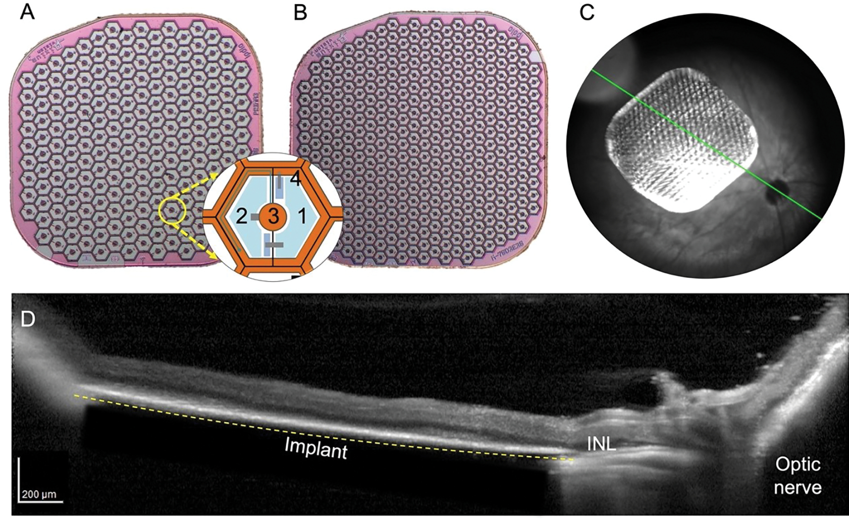
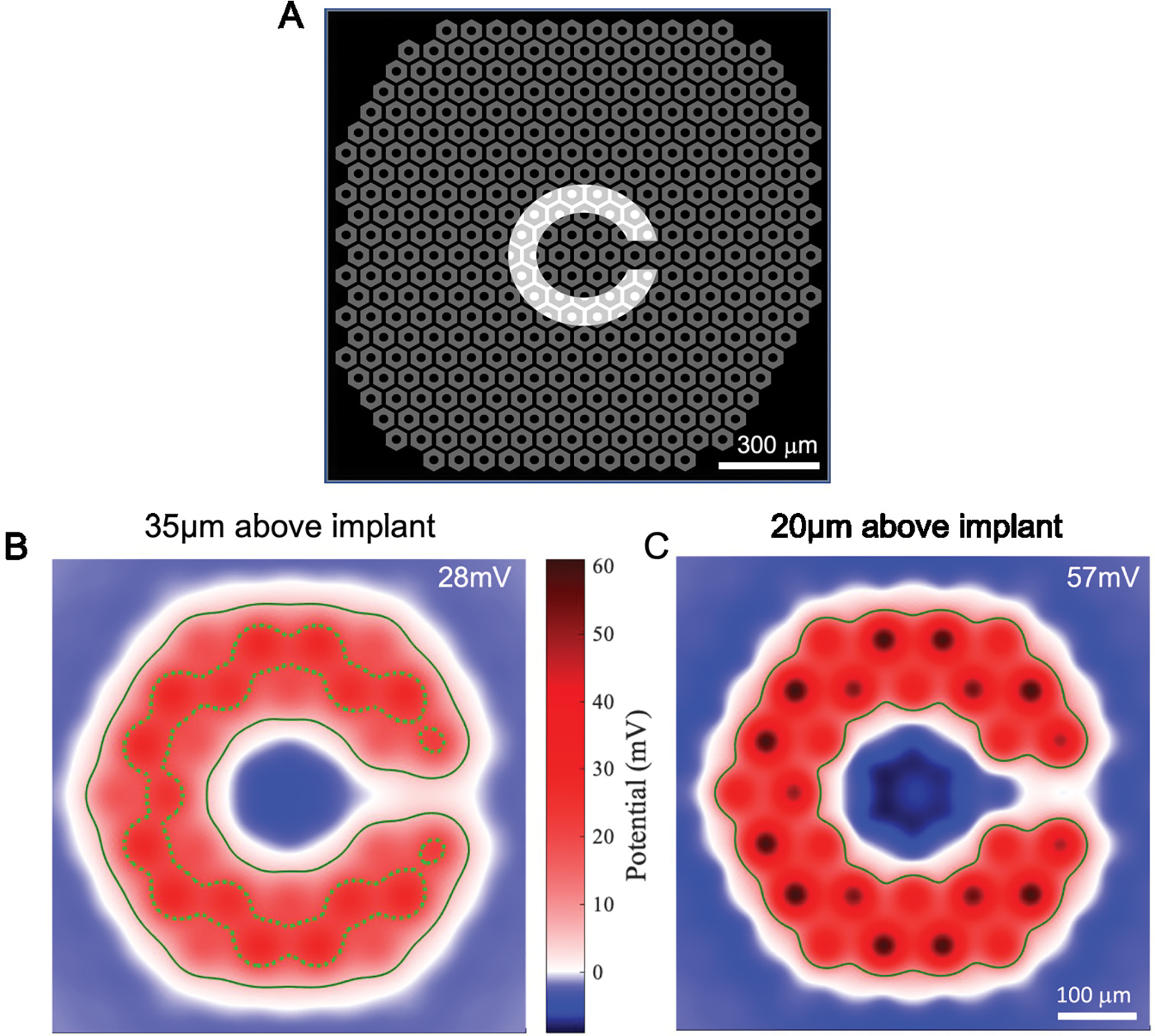
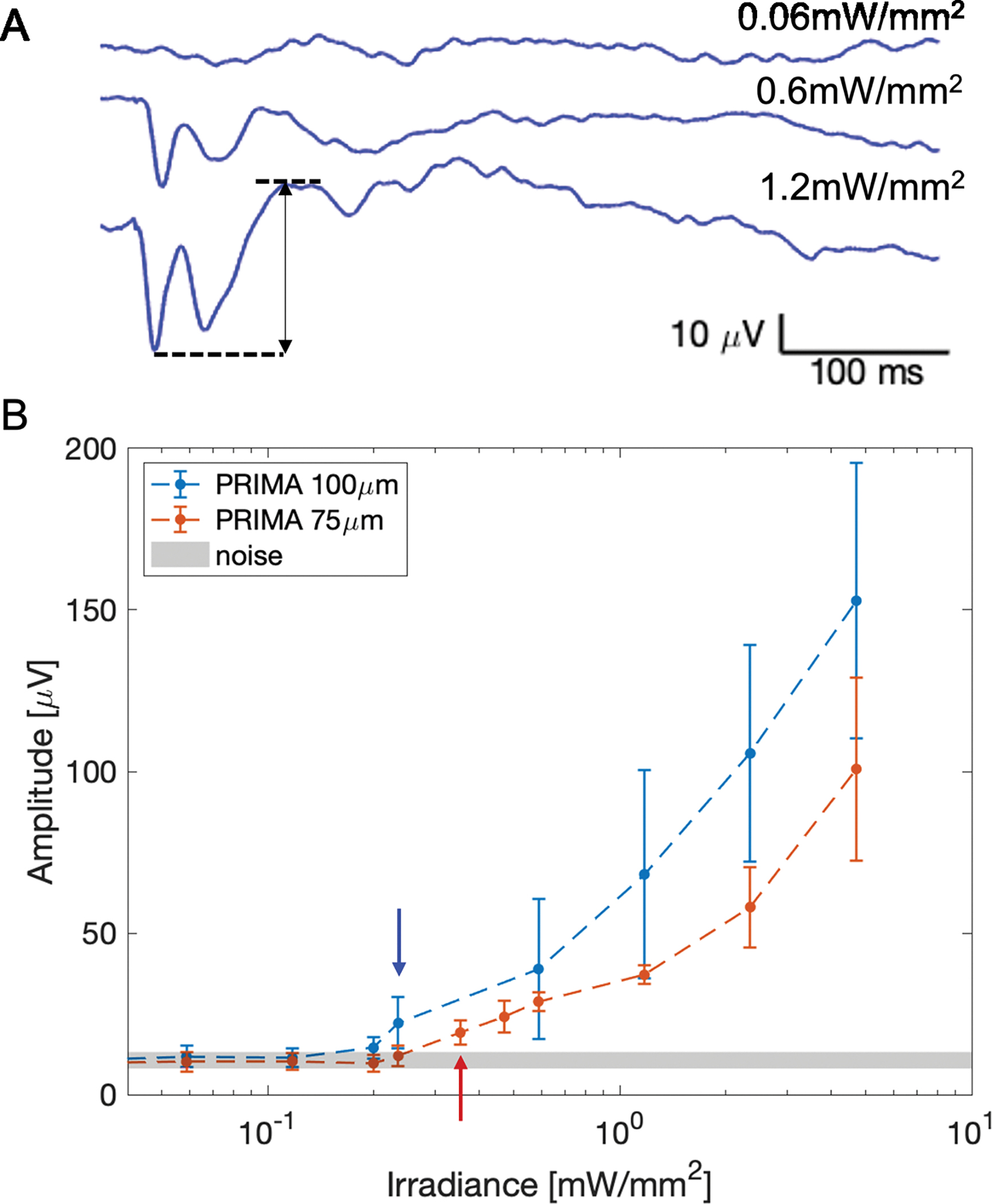
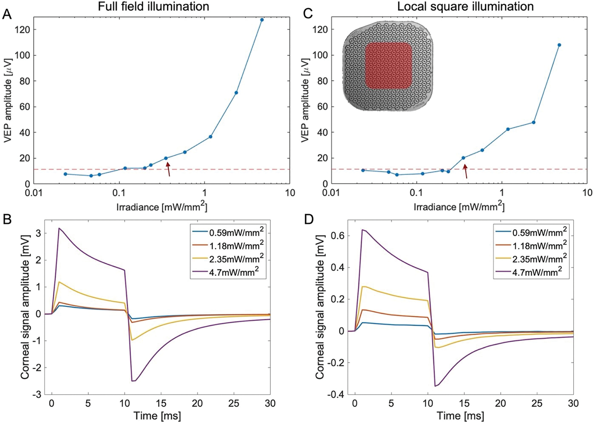
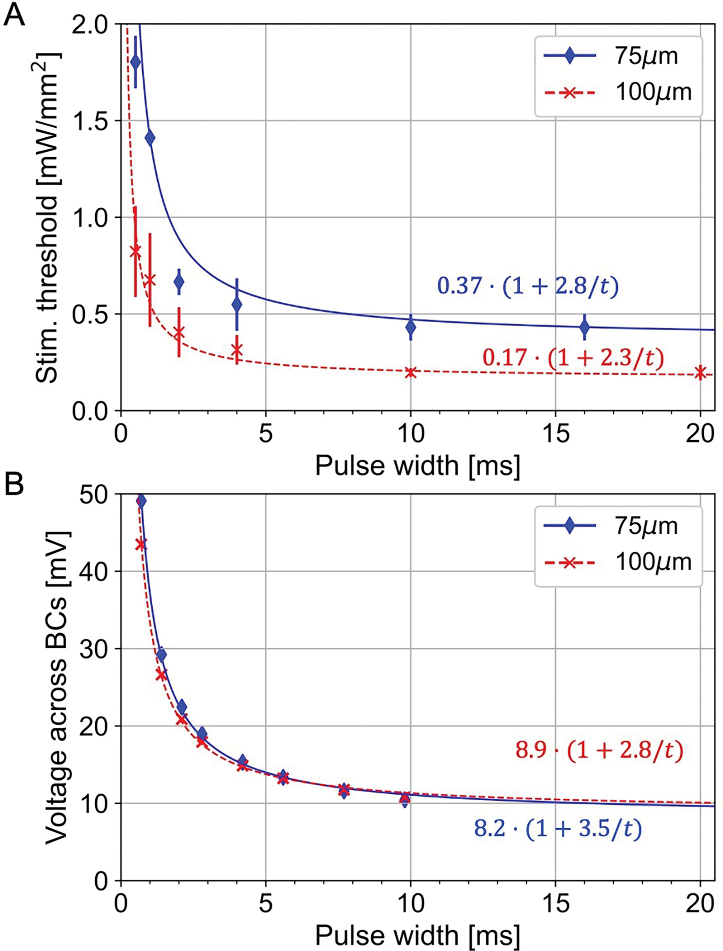
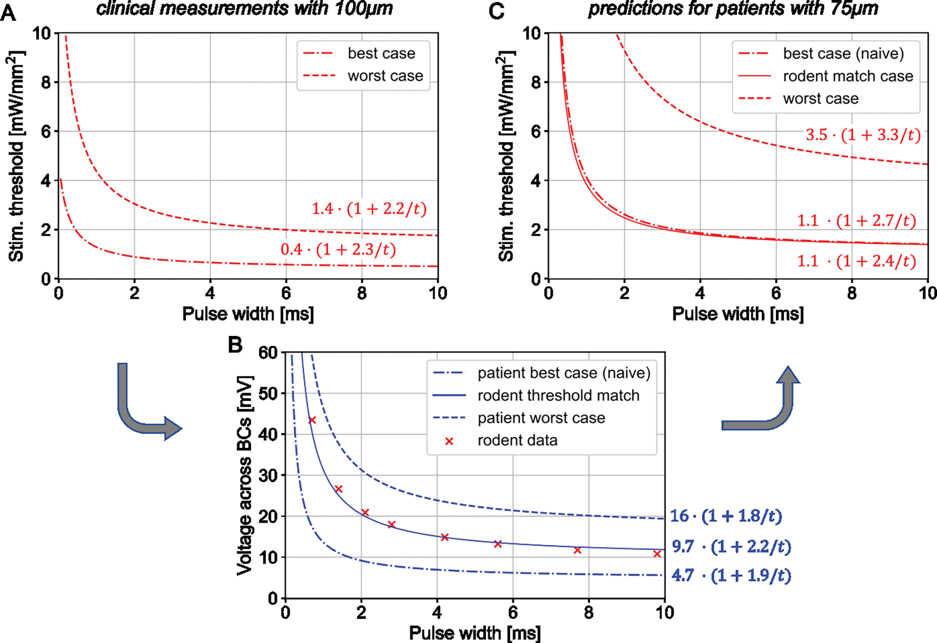
Retinal prostheses aim at restoring sight in patients with retinal degeneration by electrically stimulating the inner retinal neurons. Clinical trials with patients blinded by atrophic age-related macular degeneration using the PRIMA subretinal implant, a 2 × 2 mm array of 100m-wide photovoltaic pixels, have demonstrated a prosthetic visual acuity closely matching the pixel size. Further improvement in resolution requires smaller pixels, which, with the current bipolar design, necessitates more intense stimulation.We examine the lower limit of the pixel size for PRIMA implants by modeling the electric field, leveraging the clinical benchmarks, and using animal data to assess the stimulation strength and contrast of various patterns. Visually evoked potentials measured in Royal College of Surgeons rats with photovoltaic implants composed of 100m and 75m pixels were compared to clinical thresholds with 100m pixels. Electrical stimulation model calibrated by the clinical and rodent data was used to predict the performance of the implant with smaller pixels.PRIMA implants with 75m bipolar pixels under the maximum safe near-infrared (880 nm) illumination of 8 mW mmwith 30% duty cycle (10 ms pulses at 30 Hz) should provide a similar perceptual brightness as with 100m pixels under 3 mW mmirradiance, used in the current clinical trials. Contrast of the Landolt C pattern scaled down to 75m pixels is also similar under such illumination to that with 100m pixels, increasing the maximum acuity from 20/420 to 20/315.Computational modeling defines the minimum pixel size of the PRIMA implants as 75m. Increasing the implant width from 2 to 3 mm and reducing the pixel size from 100 to 75m will nearly quadrupole the number of pixels, which should be very beneficial for patients. Smaller pixels of the same bipolar flat geometry would require excessively intense illumination, and therefore a different pixel design should be considered for further improvement in resolution.
视网膜假体旨在通过电刺激内视网膜神经元来恢复视网膜变性患者的视力。使用 PRIMA 视网膜下植入物对因萎缩性年龄相关性黄斑变性而失明的患者进行临床试验,该植入物是一个 2×2 毫米的 100 微米宽光伏像素阵列,已证明假体视力与像素尺寸非常匹配。分辨率的进一步提高需要更小的像素,而对于当前的双极设计,这需要更强烈的刺激。我们通过建模电场、利用临床基准和使用动物数据来评估各种模式的刺激强度和对比度,来检查 PRIMA 植入物的像素尺寸下限。与具有 100 微米像素的临床阈值相比,比较了具有 100 微米和 75 微米像素的光伏植入物的皇家外科医生大鼠的视觉诱发电位。通过临床和啮齿动物数据校准的电刺激模型用于预测具有更小像素的植入物的性能。在 8 mW/mm 的最大安全近红外 (880nm) 照明下(30%占空比(30Hz 时 10ms 脉冲)),具有 75m 双极像素的 PRIMA 植入物应提供与当前临床试验中使用的 3 mW/mm 辐照度下 100m 像素相似的感知亮度。在这种照明下,缩小到 75m 像素的 Landolt C 图案对比度也与 100m 像素相似,将最大视力从 20/420 提高到 20/315。计算建模将 PRIMA 植入物的最小像素尺寸定义为 75m。将植入物宽度从 2 毫米增加到 3 毫米,并将像素尺寸从 100 微米减小到 75 微米,将使像素数量增加近四倍,这对患者非常有益。相同双极平面几何形状的更小像素将需要过于强烈的照明,因此应该考虑不同的像素设计以进一步提高分辨率。