Department of Applied Physics, Stanford University, Stanford, CA, USA.
Hansen Experimental Physics Laboratory, Stanford University, Stanford, CA, USA.
Sci Rep. 2019 Jul 23;9(1):10657. doi: 10.1038/s41598-019-47082-y.
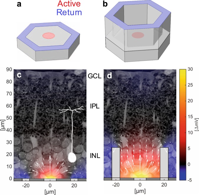
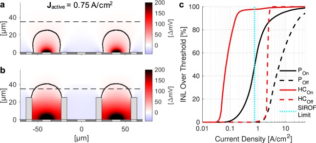
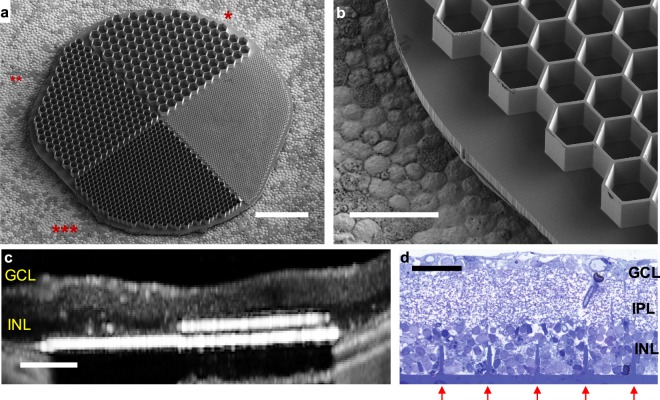
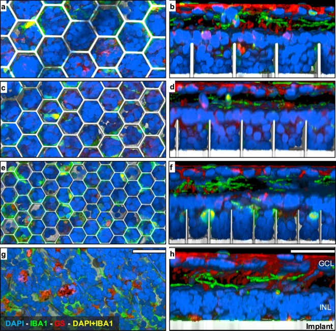
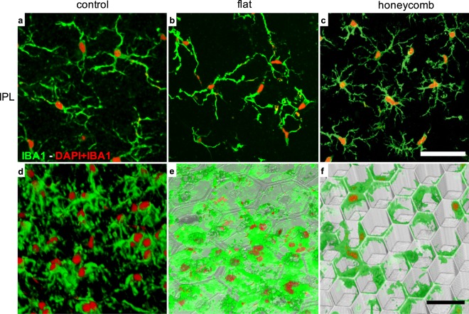
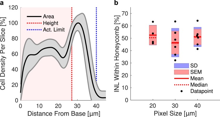
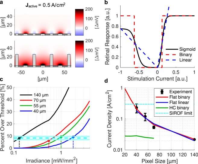
High-resolution visual prostheses require small, densely packed pixels, but limited penetration depth of the electric field formed by a planar electrode array constrains such miniaturization. We present a novel honeycomb configuration of an electrode array with vertically separated active and return electrodes designed to leverage migration of retinal cells into voids in the subretinal space. Insulating walls surrounding each pixel decouple the field penetration depth from the pixel width by aligning the electric field vertically, enabling a decrease of the pixel size down to cellular dimensions. We demonstrate that inner retinal cells migrate into the 25 μm deep honeycomb wells as narrow as 18 μm, resulting in more than half of these cells residing within the electrode cavities. Immune response to honeycombs is comparable to that with planar arrays. Modeled stimulation threshold current density with honeycombs does not increase substantially with reduced pixel size, unlike quadratic increase with planar arrays. This 3-D electrode configuration may enable functional restoration of central vision with acuity better than 20/100 for millions of patients suffering from age-related macular degeneration.
高分辨率视觉假体需要小而密集的像素,但平面电极阵列形成的电场穿透深度有限,限制了这种小型化。我们提出了一种新型的蜂窝状电极阵列结构,具有垂直分离的有源和返回电极,旨在利用视网膜细胞迁移到视网膜下空间的空隙中。每个像素周围的绝缘壁通过垂直对准电场来解耦场穿透深度和像素宽度,从而能够将像素尺寸减小到细胞尺寸。我们证明,内视网膜细胞可以迁移到深 25 μm 的蜂窝状井中,其宽度窄至 18 μm,结果超过一半的细胞位于电极腔室内。对蜂窝的免疫反应与平面阵列相当。与平面阵列的二次增加不同,用蜂窝建模的刺激阈值电流密度不会随像素尺寸的减小而显著增加。这种 3D 电极结构可以为数百万名因年龄相关性黄斑变性而导致中心视力低于 20/100 的患者实现功能恢复。