• 文献检索
  • 文档翻译
  • 深度研究
  • 学术资讯
  • Suppr Zotero 插件Zotero 插件
  • 邀请有礼
  • 套餐&价格
  • 历史记录
应用&插件
Suppr Zotero 插件Zotero 插件浏览器插件Mac 客户端Windows 客户端微信小程序
定价
高级版会员购买积分包购买API积分包
服务
文献检索文档翻译深度研究API 文档MCP 服务
关于我们
关于 Suppr公司介绍联系我们用户协议隐私条款
关注我们

Suppr 超能文献

核心技术专利:CN118964589B侵权必究
粤ICP备2023148730 号-1Suppr @ 2026

文献检索

告别复杂PubMed语法,用中文像聊天一样搜索,搜遍4000万医学文献。AI智能推荐,让科研检索更轻松。

立即免费搜索

文件翻译

保留排版,准确专业,支持PDF/Word/PPT等文件格式,支持 12+语言互译。

免费翻译文档

深度研究

AI帮你快速写综述,25分钟生成高质量综述,智能提取关键信息,辅助科研写作。

立即免费体验

使用机器学习从无对比剂心脏磁共振成像预测对比剂后信息:挑战与方法

Predicting post-contrast information from contrast agent free cardiac MRI using machine learning: Challenges and methods.

作者信息

Abdulkareem Musa, Kenawy Asmaa A, Rauseo Elisa, Lee Aaron M, Sojoudi Alireza, Amir-Khalili Alborz, Lekadir Karim, Young Alistair A, Barnes Michael R, Barckow Philipp, Khanji Mohammed Y, Aung Nay, Petersen Steffen E

机构信息

Barts Heart Centre, Barts Health National Health Service (NHS) Trust, London, United Kingdom.

National Institute for Health Research (NIHR) Barts Biomedical Research Centre, William Harvey Research Institute, Queen Mary University of London, London, United Kingdom.

出版信息

Front Cardiovasc Med. 2022 Jul 27;9:894503. doi: 10.3389/fcvm.2022.894503. eCollection 2022.

DOI:10.3389/fcvm.2022.894503
PMID:36051279
原文链接:https://pmc.ncbi.nlm.nih.gov/articles/PMC9426684/
Abstract

OBJECTIVES

Currently, administering contrast agents is necessary for accurately visualizing and quantifying presence, location, and extent of myocardial infarction (MI) with cardiac magnetic resonance (CMR). In this study, our objective is to investigate and analyze pre- and post-contrast CMR images with the goal of predicting post-contrast information using pre-contrast information only. We propose methods and identify challenges.

METHODS

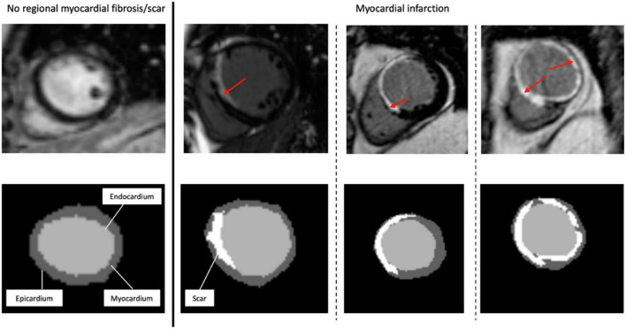
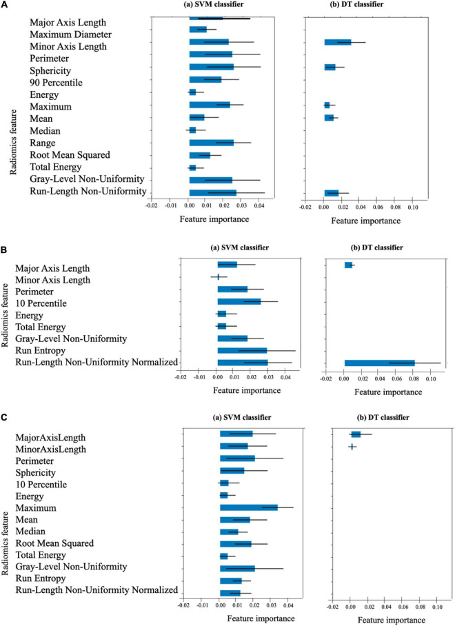
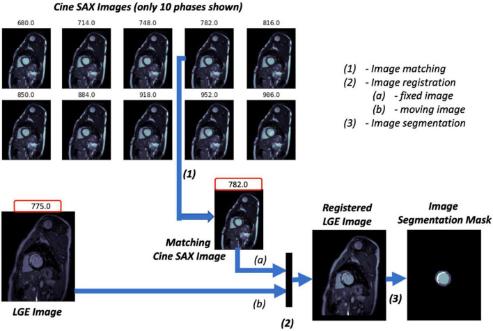
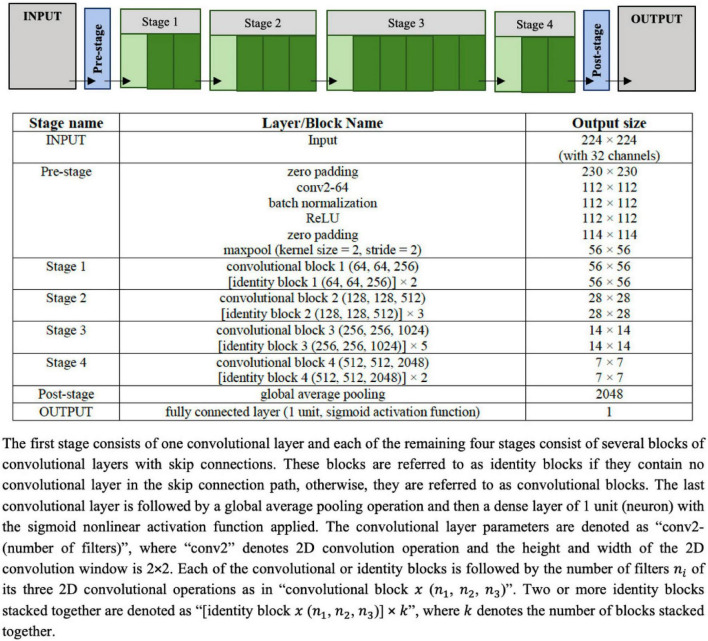
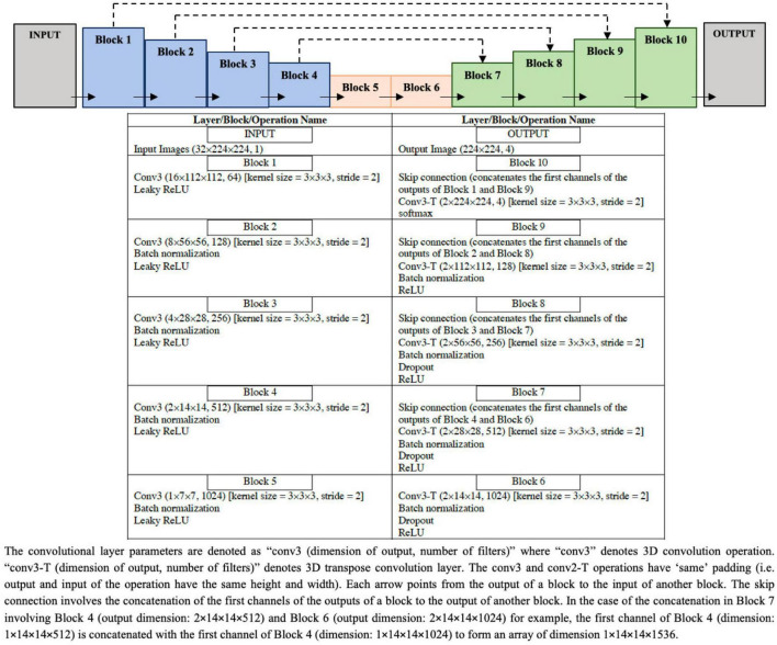
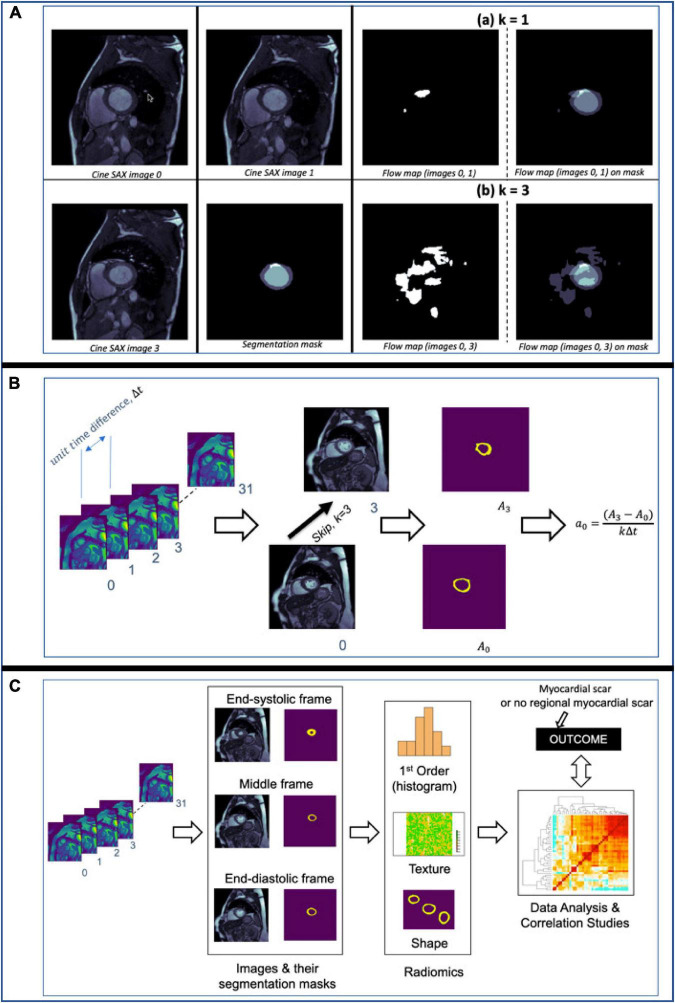
The study population consists of 272 retrospectively selected CMR studies with diagnoses of MI ( = 108) and healthy controls ( = 164). We describe a pipeline for pre-processing this dataset for analysis. After data feature engineering, 722 cine short-axis (SAX) images and segmentation mask pairs were used for experimentation. This constitutes 506, 108, and 108 pairs for the training, validation, and testing sets, respectively. We use deep learning (DL) segmentation (UNet) and classification (ResNet50) models to discover the extent and location of the scar and classify between the ischemic cases and healthy cases (i.e., cases with no regional myocardial scar) from the pre-contrast cine SAX image frames, respectively. We then capture complex data patterns that represent subtle signal and functional changes in the cine SAX images due to MI using optical flow, rate of change of myocardial area, and radiomics data. We apply this dataset to explore two supervised learning methods, namely, the support vector machines (SVM) and the decision tree (DT) methods, to develop predictive models for classifying pre-contrast cine SAX images as being a case of MI or healthy.

RESULTS

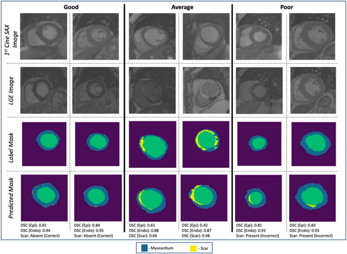
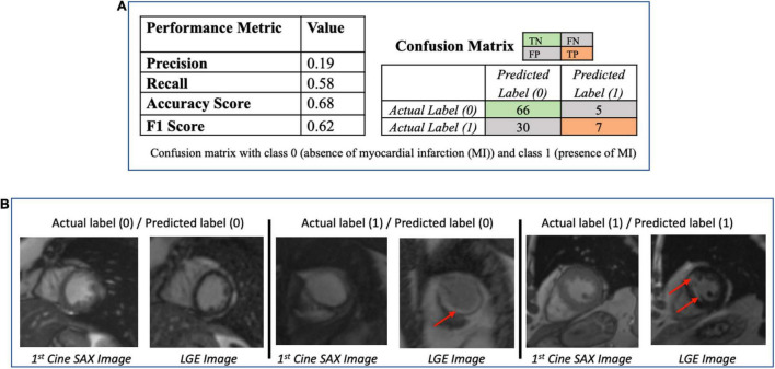
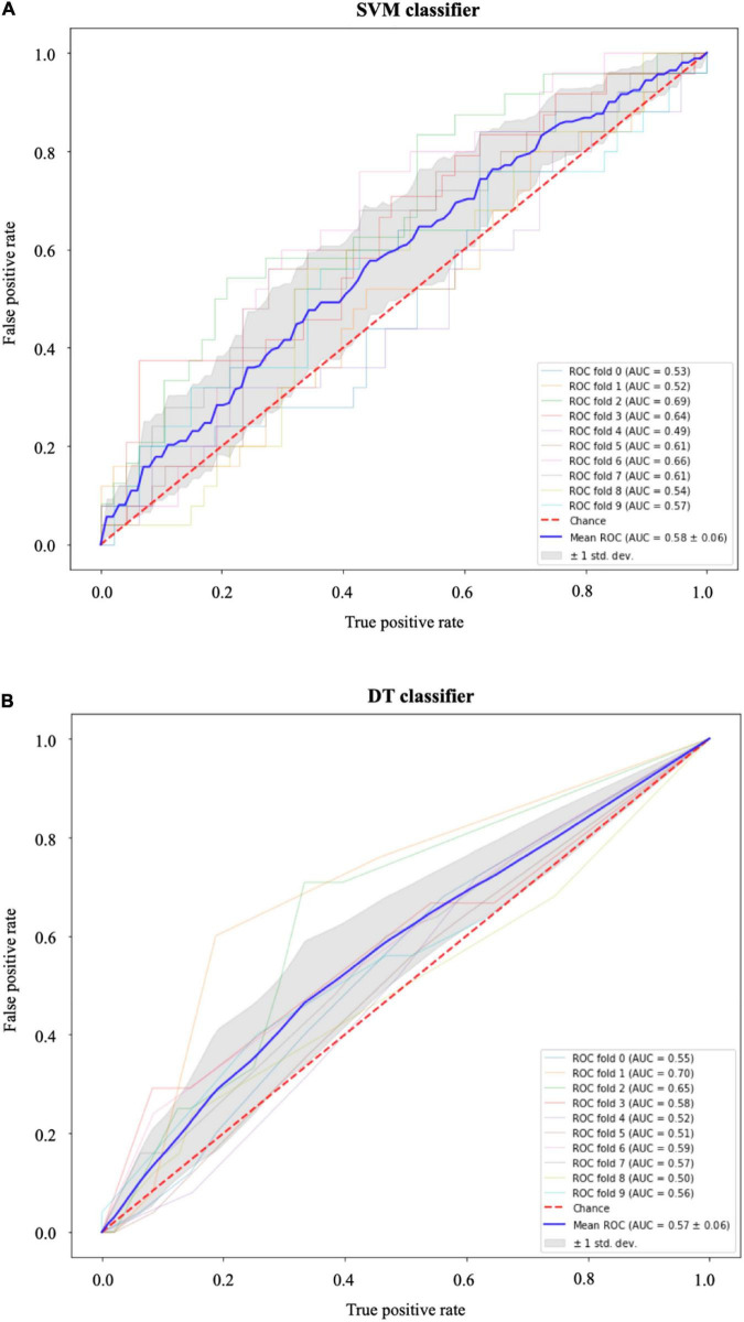
Overall, for the UNet segmentation model, the performance based on the mean Dice score for the test set ( = 108) is 0.75 (±0.20) for the endocardium, 0.51 (±0.21) for the epicardium and 0.20 (±0.17) for the scar. For the classification task, the accuracy, F1 and precision scores of 0.68, 0.69, and 0.64, respectively, were achieved with the SVM model, and of 0.62, 0.63, and 0.72, respectively, with the DT model.

CONCLUSION

We have presented some promising approaches involving DL, SVM, and DT methods in an attempt to accurately predict contrast information from non-contrast images. While our initial results are modest for this challenging task, this area of research still poses several open problems.

摘要

目的

目前,使用心脏磁共振成像(CMR)准确显示和量化心肌梗死(MI)的存在、位置及范围时,注射造影剂是必要的。在本研究中,我们的目的是研究和分析注射造影剂前后的CMR图像,目标是仅使用注射造影剂前的信息来预测注射造影剂后的信息。我们提出了方法并识别了挑战。

方法

研究人群包括272例经回顾性选择的CMR研究,其中诊断为MI的有108例,健康对照有164例。我们描述了一个用于预处理此数据集以进行分析的流程。经过数据特征工程后,722对电影短轴(SAX)图像和分割掩码对用于实验。这分别构成了训练集、验证集和测试集的506对、108对和108对。我们使用深度学习(DL)分割(UNet)和分类(ResNet50)模型分别从注射造影剂前的电影SAX图像帧中发现瘢痕的范围和位置,并对缺血病例和健康病例(即无局部心肌瘢痕的病例)进行分类。然后,我们使用光流、心肌面积变化率和放射组学数据来捕捉代表由于MI导致的电影SAX图像中细微信号和功能变化的复杂数据模式。我们应用此数据集探索两种监督学习方法,即支持向量机(SVM)和决策树(DT)方法,以开发用于将注射造影剂前的电影SAX图像分类为MI病例或健康病例的预测模型。

结果

总体而言,对于UNet分割模型,测试集(n = 108)的基于平均Dice分数的性能,心内膜为0.75(±0.20),心外膜为0.51(±0.21),瘢痕为0.20(±0.17)。对于分类任务,SVM模型的准确率、F1分数和精确率分别为0.68、0.69和0.64,DT模型的分别为0.62、0.63和0.72。

结论

我们提出了一些涉及DL、SVM和DT方法的有前景的方法,试图从非对比图像中准确预测对比信息。虽然我们针对这项具有挑战性的任务的初步结果并不理想,但该研究领域仍然存在几个未解决的问题。

https://cdn.ncbi.nlm.nih.gov/pmc/blobs/ad9d/9426684/a80b2aa9f92a/fcvm-09-894503-g010.jpg
https://cdn.ncbi.nlm.nih.gov/pmc/blobs/ad9d/9426684/1f5f45adb811/fcvm-09-894503-g001.jpg
https://cdn.ncbi.nlm.nih.gov/pmc/blobs/ad9d/9426684/86e3c7f86d76/fcvm-09-894503-g002.jpg
https://cdn.ncbi.nlm.nih.gov/pmc/blobs/ad9d/9426684/cabc80c580a2/fcvm-09-894503-g003.jpg
https://cdn.ncbi.nlm.nih.gov/pmc/blobs/ad9d/9426684/ac26fe121de9/fcvm-09-894503-g004.jpg
https://cdn.ncbi.nlm.nih.gov/pmc/blobs/ad9d/9426684/859b70a0c5e8/fcvm-09-894503-g005.jpg
https://cdn.ncbi.nlm.nih.gov/pmc/blobs/ad9d/9426684/45a418a3ec91/fcvm-09-894503-g006.jpg
https://cdn.ncbi.nlm.nih.gov/pmc/blobs/ad9d/9426684/a6dabf628955/fcvm-09-894503-g007.jpg
https://cdn.ncbi.nlm.nih.gov/pmc/blobs/ad9d/9426684/ad8d4016be06/fcvm-09-894503-g008.jpg
https://cdn.ncbi.nlm.nih.gov/pmc/blobs/ad9d/9426684/8ec3a973c2b0/fcvm-09-894503-g009.jpg
https://cdn.ncbi.nlm.nih.gov/pmc/blobs/ad9d/9426684/a80b2aa9f92a/fcvm-09-894503-g010.jpg

相似文献

1
Predicting post-contrast information from contrast agent free cardiac MRI using machine learning: Challenges and methods.使用机器学习从无对比剂心脏磁共振成像预测对比剂后信息:挑战与方法
Front Cardiovasc Med. 2022 Jul 27;9:894503. doi: 10.3389/fcvm.2022.894503. eCollection 2022.
2
Non-contrast Cine Cardiac Magnetic Resonance image radiomics features and machine learning algorithms for myocardial infarction detection.用于心肌梗死检测的非对比动态心脏磁共振图像放射组学特征及机器学习算法
Comput Biol Med. 2022 Feb;141:105145. doi: 10.1016/j.compbiomed.2021.105145. Epub 2021 Dec 15.
3
Radiomics-based detection of acute myocardial infarction on noncontrast enhanced midventricular short-axis cine CMR images.基于影像组学的非增强心室中部短轴电影心脏磁共振成像检测急性心肌梗死
Int J Cardiovasc Imaging. 2024 Jun;40(6):1211-1220. doi: 10.1007/s10554-024-03089-9. Epub 2024 Apr 17.
4
Joint Deep Learning Framework for Image Registration and Segmentation of Late Gadolinium Enhanced MRI and Cine Cardiac MRI.用于延迟钆增强磁共振成像和心脏电影磁共振成像的图像配准与分割的联合深度学习框架
Proc SPIE Int Soc Opt Eng. 2021 Feb;11598. doi: 10.1117/12.2581386. Epub 2021 Feb 15.
5
Automatic cardiac cine MRI segmentation and heart disease classification.自动心脏电影磁共振成像分割与心脏病分类。
Comput Med Imaging Graph. 2021 Mar;88:101864. doi: 10.1016/j.compmedimag.2021.101864. Epub 2021 Jan 13.
6
Automated left and right ventricular chamber segmentation in cardiac magnetic resonance images using dense fully convolutional neural network.使用密集全卷积神经网络对心脏磁共振图像进行自动左右心室腔分割
Comput Methods Programs Biomed. 2021 Jun;204:106059. doi: 10.1016/j.cmpb.2021.106059. Epub 2021 Mar 21.
7
Differentiating between cardiac amyloidosis and hypertrophic cardiomyopathy on non-contrast cine-magnetic resonance images using machine learning-based radiomics.使用基于机器学习的放射组学在非对比电影磁共振图像上鉴别心脏淀粉样变性和肥厚型心肌病。
Front Cardiovasc Med. 2022 Oct 26;9:1001269. doi: 10.3389/fcvm.2022.1001269. eCollection 2022.
8
Cardiac magnetic resonance image-based classification of the risk of arrhythmias in post-myocardial infarction patients.基于心脏磁共振成像的心肌梗死后患者心律失常风险分类
Artif Intell Med. 2015 Jul;64(3):205-15. doi: 10.1016/j.artmed.2015.06.001. Epub 2015 Jul 4.
9
New Imaging Signatures of Cardiac Alterations in Ischaemic Heart Disease and Cerebrovascular Disease Using CMR Radiomics.使用心脏磁共振成像放射组学技术对缺血性心脏病和脑血管病中心脏改变的新成像特征
Front Cardiovasc Med. 2021 Sep 23;8:716577. doi: 10.3389/fcvm.2021.716577. eCollection 2021.
10
Ensemble learning of myocardial displacements for myocardial infarction detection in echocardiography.用于超声心动图中心肌梗死检测的心肌位移集成学习
Front Cardiovasc Med. 2023 Oct 13;10:1185172. doi: 10.3389/fcvm.2023.1185172. eCollection 2023.

引用本文的文献

1
An Innovative Artificial Intelligence Classification Model for Non-Ischemic Cardiomyopathy Utilizing Cardiac Biomechanics Derived from Magnetic Resonance Imaging.一种利用磁共振成像衍生的心脏生物力学的非缺血性心肌病创新人工智能分类模型。
Bioengineering (Basel). 2025 Jun 19;12(6):670. doi: 10.3390/bioengineering12060670.
2
Toward understanding the balanced steady-state free precession signal intensity changes in cine cardiac magnetic resonance imaging: A preliminary evaluation in healthy subjects pre- and postcontrast.关于理解心脏电影磁共振成像中平衡稳态自由进动信号强度变化:对比剂注射前后健康受试者的初步评估
J Cardiovasc Magn Reson. 2025 May 8;27(2):101908. doi: 10.1016/j.jocmr.2025.101908.
3

本文引用的文献

1
Cardiac segmentation on late gadolinium enhancement MRI: A benchmark study from multi-sequence cardiac MR segmentation challenge.延迟钆增强磁共振成像的心脏分割:来自多序列心脏磁共振分割挑战赛的一项基准研究。
Med Image Anal. 2022 Oct;81:102528. doi: 10.1016/j.media.2022.102528. Epub 2022 Jul 9.
2
Generalizable Framework for Atrial Volume Estimation for Cardiac CT Images Using Deep Learning With Quality Control Assessment.基于深度学习和质量控制评估的心脏CT图像心房容积估计通用框架
Front Cardiovasc Med. 2022 Jan 28;9:822269. doi: 10.3389/fcvm.2022.822269. eCollection 2022.
3
Non-contrast Cine Cardiac Magnetic Resonance image radiomics features and machine learning algorithms for myocardial infarction detection.
Machine learning-based interpretation of non-contrast feature tracking strain analysis and T1/T2 mapping for assessing myocardial viability.
基于机器学习的非对比特征追踪应变分析及T1/T2映射用于评估心肌存活的解读
Sci Rep. 2025 Jan 4;15(1):753. doi: 10.1038/s41598-024-85029-0.
4
AI-powered contrast-free cardiovascular magnetic resonance imaging for myocardial infarction.用于心肌梗死的人工智能无对比剂心血管磁共振成像
Front Cardiovasc Med. 2024 Nov 21;11:1457498. doi: 10.3389/fcvm.2024.1457498. eCollection 2024.
5
Imaging for the diagnosis of acute myocarditis: can artificial intelligence improve diagnostic performance?用于急性心肌炎诊断的影像学检查:人工智能能否提高诊断效能?
Front Cardiovasc Med. 2024 Aug 29;11:1408574. doi: 10.3389/fcvm.2024.1408574. eCollection 2024.
6
Quantification of Epicardial Adipose Tissue Volume and Attenuation for Cardiac CT Scans Using Deep Learning in a Single Multi-Task Framework.在单一多任务框架中使用深度学习对心脏CT扫描的心外膜脂肪组织体积和衰减进行量化。
Rev Cardiovasc Med. 2022 Dec 20;23(12):412. doi: 10.31083/j.rcm2312412. eCollection 2022 Dec.
7
Radiomics-based detection of acute myocardial infarction on noncontrast enhanced midventricular short-axis cine CMR images.基于影像组学的非增强心室中部短轴电影心脏磁共振成像检测急性心肌梗死
Int J Cardiovasc Imaging. 2024 Jun;40(6):1211-1220. doi: 10.1007/s10554-024-03089-9. Epub 2024 Apr 17.
8
Artificial Intelligence Applications in Cardiovascular Magnetic Resonance Imaging: Are We on the Path to Avoiding the Administration of Contrast Media?人工智能在心血管磁共振成像中的应用:我们是否正朝着避免使用造影剂的方向发展?
Diagnostics (Basel). 2023 Jun 14;13(12):2061. doi: 10.3390/diagnostics13122061.
用于心肌梗死检测的非对比动态心脏磁共振图像放射组学特征及机器学习算法
Comput Biol Med. 2022 Feb;141:105145. doi: 10.1016/j.compbiomed.2021.105145. Epub 2021 Dec 15.
4
New Imaging Signatures of Cardiac Alterations in Ischaemic Heart Disease and Cerebrovascular Disease Using CMR Radiomics.使用心脏磁共振成像放射组学技术对缺血性心脏病和脑血管病中心脏改变的新成像特征
Front Cardiovasc Med. 2021 Sep 23;8:716577. doi: 10.3389/fcvm.2021.716577. eCollection 2021.
5
Automated Noncontrast Myocardial Tissue Characterization for Hypertrophic Cardiomyopathy: Holy Grail or False Prophet?肥厚型心肌病的自动非对比心肌组织特征分析:圣杯还是虚假预言?
Circulation. 2021 Aug 24;144(8):600-603. doi: 10.1161/CIRCULATIONAHA.121.055791. Epub 2021 Aug 23.
6
Recent Advances in Fibrosis and Scar Segmentation From Cardiac MRI: A State-of-the-Art Review and Future Perspectives.心脏磁共振成像中纤维化和瘢痕分割的最新进展:现状综述与未来展望
Front Physiol. 2021 Aug 3;12:709230. doi: 10.3389/fphys.2021.709230. eCollection 2021.
7
Automated Left Ventricle Ischemic Scar Detection in CT Using Deep Neural Networks.使用深度神经网络在CT中自动检测左心室缺血性瘢痕
Front Cardiovasc Med. 2021 Jul 2;8:655252. doi: 10.3389/fcvm.2021.655252. eCollection 2021.
8
Toward Replacing Late Gadolinium Enhancement With Artificial Intelligence Virtual Native Enhancement for Gadolinium-Free Cardiovascular Magnetic Resonance Tissue Characterization in Hypertrophic Cardiomyopathy.用人工智能虚拟原生增强替代晚期钆增强,实现肥厚型心肌病的无钆心血管磁共振组织特征分析。
Circulation. 2021 Aug 24;144(8):589-599. doi: 10.1161/CIRCULATIONAHA.121.054432. Epub 2021 Jul 7.
9
The Promise of AI in Detection, Diagnosis, and Epidemiology for Combating COVID-19: Beyond the Hype.人工智能在抗击新冠疫情中的检测、诊断和流行病学方面的前景:超越炒作。
Front Artif Intell. 2021 May 14;4:652669. doi: 10.3389/frai.2021.652669. eCollection 2021.
10
Automated Segmental Analysis of Fully Quantitative Myocardial Blood Flow Maps by First-Pass Perfusion Cardiovascular Magnetic Resonance.通过首次通过灌注心血管磁共振对全定量心肌血流图进行自动节段分析。
IEEE Access. 2021;9:52796-52811. doi: 10.1109/access.2021.3070320. Epub 2021 Apr 1.