• 文献检索
  • 文档翻译
  • 深度研究
  • 学术资讯
  • Suppr Zotero 插件Zotero 插件
  • 邀请有礼
  • 套餐&价格
  • 历史记录
应用&插件
Suppr Zotero 插件Zotero 插件浏览器插件Mac 客户端Windows 客户端微信小程序
定价
高级版会员购买积分包购买API积分包
服务
文献检索文档翻译深度研究API 文档MCP 服务
关于我们
关于 Suppr公司介绍联系我们用户协议隐私条款
关注我们

Suppr 超能文献

核心技术专利:CN118964589B侵权必究
粤ICP备2023148730 号-1Suppr @ 2026

文献检索

告别复杂PubMed语法,用中文像聊天一样搜索,搜遍4000万医学文献。AI智能推荐,让科研检索更轻松。

立即免费搜索

文件翻译

保留排版,准确专业,支持PDF/Word/PPT等文件格式,支持 12+语言互译。

免费翻译文档

深度研究

AI帮你快速写综述,25分钟生成高质量综述,智能提取关键信息,辅助科研写作。

立即免费体验

用于延迟钆增强磁共振成像和心脏电影磁共振成像的图像配准与分割的联合深度学习框架

Joint Deep Learning Framework for Image Registration and Segmentation of Late Gadolinium Enhanced MRI and Cine Cardiac MRI.

作者信息

Upendra Roshan Reddy, Simon Richard, Linte Cristian A

机构信息

Center for Imaging Science, Rochester Institute of Technology, Rochester, NY, USA.

Biomedical Engineering, Rochester Institute of Technology, Rochester, NY, USA.

出版信息

Proc SPIE Int Soc Opt Eng. 2021 Feb;11598. doi: 10.1117/12.2581386. Epub 2021 Feb 15.

DOI:10.1117/12.2581386
PMID:34079155
原文链接:https://pmc.ncbi.nlm.nih.gov/articles/PMC8168979/
Abstract

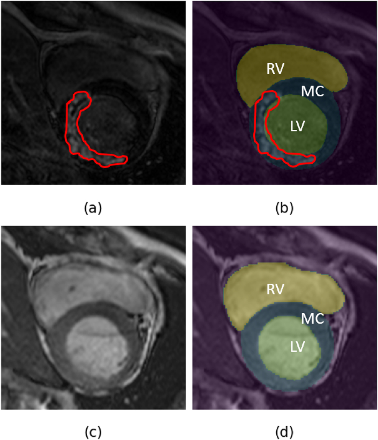
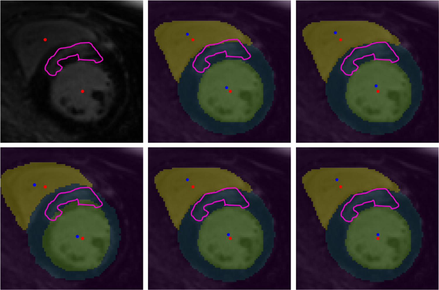
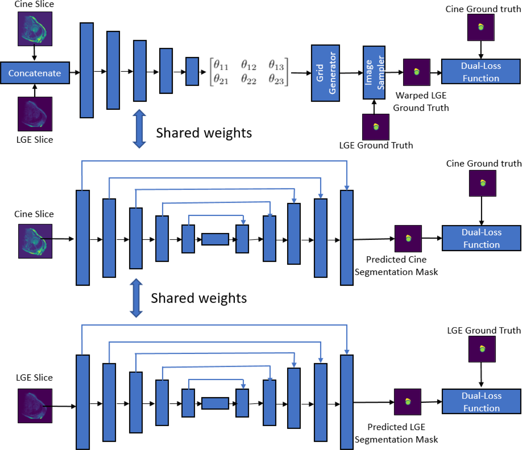
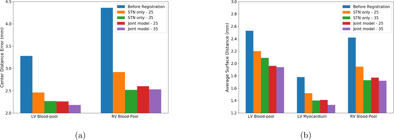
Late gadolinium enhanced (LGE) cardiac magnetic resonance (CMR) imaging, the current benchmark for assessment of myocardium viability, enables the identification and quantification of the compromised myocardial tissue regions, as they appear hyper-enhanced compared to the surrounding, healthy myocardium. However, in LGE CMR images, the reduced contrast between the left ventricle (LV) myocardium and LV blood-pool hampers the accurate delineation of the LV myocardium. On the other hand, the balanced-Steady State Free Precession (bSSFP) cine CMR imaging provides high resolution images ideal for accurate segmentation of the cardiac chambers. In the interest of generating patient-specific hybrid 3D and 4D anatomical models of the heart, to identify and quantify the compromised myocardial tissue regions for revascularization therapy planning, in our previous work, we presented a spatial transformer network (STN) based convolutional neural network (CNN) architecture for registration of LGE and bSSFP cine CMR image datasets made available through the 2019 Multi-Sequence Cardiac Magnetic Resonance Segmentation Challenge (MS-CMRSeg). We performed a supervised registration by leveraging the region of interest (RoI) information using the manual annotations of the LV blood-pool, LV myocardium and right ventricle (RV) blood-pool provided for both the LGE and the bSSFP cine CMR images. In order to reduce the reliance on the number of manual annotations for training such network, we propose a joint deep learning framework consisting of three branches: a STN based RoI guided CNN for registration of LGE and bSSFP cine CMR images, an U-Net model for segmentation of bSSFP cine CMR images, and an U-Net model for segmentation of LGE CMR images. This results in learning of a joint multi-scale feature encoder by optimizing all three branches of the network architecture simultaneously. Our experiments show that the registration results obtained by training 25 of the available 45 image datasets in a joint deep learning framework is comparable to the registration results obtained by stand-alone STN based CNN model by training 35 of the available 45 image datasets and also shows significant improvement in registration performance when compared to the results achieved by the stand-alone STN based CNN model by training 25 of the available 45 image datasets.

摘要

延迟钆增强(LGE)心脏磁共振(CMR)成像作为目前评估心肌活力的基准,能够识别和量化受损心肌组织区域,因为这些区域与周围健康心肌相比呈现高增强。然而,在LGE CMR图像中,左心室(LV)心肌与LV血池之间对比度降低,妨碍了LV心肌的准确勾画。另一方面,平衡稳态自由进动(bSSFP)电影CMR成像提供了高分辨率图像,非常适合心脏腔室的准确分割。为了生成针对患者的心脏三维和四维解剖模型,以识别和量化受损心肌组织区域用于血运重建治疗规划,在我们之前的工作中,我们提出了一种基于空间变换网络(STN)的卷积神经网络(CNN)架构,用于对通过2019年多序列心脏磁共振分割挑战赛(MS-CMRSeg)提供的LGE和bSSFP电影CMR图像数据集进行配准。我们通过利用左心室血池、左心室心肌和右心室(RV)血池的手动标注所提供的感兴趣区域(RoI)信息来进行监督配准,这些标注同时提供给LGE和bSSFP电影CMR图像。为了减少训练此类网络对手动标注数量的依赖,我们提出了一个联合深度学习框架,该框架由三个分支组成:一个基于STN的RoI引导CNN用于LGE和bSSFP电影CMR图像的配准,一个U-Net模型用于bSSFP电影CMR图像的分割,以及一个U-Net模型用于LGE CMR图像的分割。这通过同时优化网络架构的所有三个分支,实现了联合多尺度特征编码器的学习。我们的实验表明,在联合深度学习框架中训练45个可用图像数据集中的25个所获得的配准结果,与通过训练45个可用图像数据集中的35个基于独立STN的CNN模型所获得的配准结果相当,并且与通过训练45个可用图像数据集中的25个基于独立STN的CNN模型所取得的结果相比,在配准性能上有显著提升。

https://cdn.ncbi.nlm.nih.gov/pmc/blobs/5893/8168979/ec0f9e3b2a5b/nihms-1705229-f0004.jpg
https://cdn.ncbi.nlm.nih.gov/pmc/blobs/5893/8168979/cab7ac63ee14/nihms-1705229-f0001.jpg
https://cdn.ncbi.nlm.nih.gov/pmc/blobs/5893/8168979/1ee07c6d4ca2/nihms-1705229-f0002.jpg
https://cdn.ncbi.nlm.nih.gov/pmc/blobs/5893/8168979/505a20cb6763/nihms-1705229-f0003.jpg
https://cdn.ncbi.nlm.nih.gov/pmc/blobs/5893/8168979/ec0f9e3b2a5b/nihms-1705229-f0004.jpg

相似文献

1
Joint Deep Learning Framework for Image Registration and Segmentation of Late Gadolinium Enhanced MRI and Cine Cardiac MRI.用于延迟钆增强磁共振成像和心脏电影磁共振成像的图像配准与分割的联合深度学习框架
Proc SPIE Int Soc Opt Eng. 2021 Feb;11598. doi: 10.1117/12.2581386. Epub 2021 Feb 15.
2
A Supervised Image Registration Approach for Late Gadolinium Enhanced MRI and Cine Cardiac MRI Using Convolutional Neural Networks.一种使用卷积神经网络的用于延迟钆增强磁共振成像和电影心脏磁共振成像的监督图像配准方法。
Med Image Underst Anal. 2020 Jul;1248:208-220. doi: 10.1007/978-3-030-52791-4_17. Epub 2020 Jul 8.
3
Improved Quantification of Myocardium Scar in Late Gadolinium Enhancement Images: Deep Learning Based Image Fusion Approach.基于深度学习的图像融合方法提高钆延迟增强图像中心肌瘢痕的定量评估。
J Magn Reson Imaging. 2021 Jul;54(1):303-312. doi: 10.1002/jmri.27555. Epub 2021 Feb 17.
4
Evaluation of fully automated myocardial segmentation techniques in native and contrast-enhanced T1-mapping cardiovascular magnetic resonance images using fully convolutional neural networks.使用全卷积神经网络评估 native 和 contrast-enhanced T1-mapping 心血管磁共振成像中的全自动心肌分割技术。
Med Phys. 2021 Jan;48(1):215-226. doi: 10.1002/mp.14574. Epub 2020 Dec 1.
5
Segmentation of left ventricle in late gadolinium enhanced MRI through 2D-4D registration for infarct localization in 3D patient-specific left ventricular model.通过二维到四维配准对晚期钆增强 MRI 中的左心室进行分段,以便在三维患者特定左心室模型中定位梗死部位。
Magn Reson Med. 2019 Feb;81(2):1385-1398. doi: 10.1002/mrm.27486. Epub 2018 Sep 19.
6
Anatomically informed deep learning on contrast-enhanced cardiac magnetic resonance imaging for scar segmentation and clinical feature extraction.基于解剖学信息的深度学习在对比增强心脏磁共振成像上用于瘢痕分割和临床特征提取
Cardiovasc Digit Health J. 2021 Nov 26;3(1):2-13. doi: 10.1016/j.cvdhj.2021.11.007. eCollection 2022 Feb.
7
Deep learning architecture for 3D image super-resolution of late gadolinium enhanced cardiac MRI.用于延迟钆增强心脏磁共振成像3D图像超分辨率的深度学习架构。
J Med Imaging (Bellingham). 2023 Sep;10(5):051808. doi: 10.1117/1.JMI.10.5.051808. Epub 2023 May 24.
8
A distance map regularized CNN for cardiac cine MR image segmentation.基于距离图正则化卷积神经网络的心电影磁共振图像分割。
Med Phys. 2019 Dec;46(12):5637-5651. doi: 10.1002/mp.13853. Epub 2019 Oct 31.
9
Cardiac segmentation on late gadolinium enhancement MRI: A benchmark study from multi-sequence cardiac MR segmentation challenge.延迟钆增强磁共振成像的心脏分割:来自多序列心脏磁共振分割挑战赛的一项基准研究。
Med Image Anal. 2022 Oct;81:102528. doi: 10.1016/j.media.2022.102528. Epub 2022 Jul 9.
10
Automated Segmentation of Cardiac Chambers from Cine Cardiac MRI Using an Adversarial Network Architecture.使用对抗网络架构从心脏电影磁共振成像中自动分割心脏腔室
Proc SPIE Int Soc Opt Eng. 2020 Feb;11315. doi: 10.1117/12.2550656. Epub 2020 Mar 16.

引用本文的文献

1
Improving the efficiency and accuracy of cardiovascular magnetic resonance with artificial intelligence-review of evidence and proposition of a roadmap to clinical translation.利用人工智能提高心血管磁共振成像的效率和准确性——证据综述及临床转化路线图建议
J Cardiovasc Magn Reson. 2024;26(2):101051. doi: 10.1016/j.jocmr.2024.101051. Epub 2024 Jun 22.
2
Medical image alignment based on landmark- and approximate contour-matching.基于地标和近似轮廓匹配的医学图像配准
J Med Imaging (Bellingham). 2021 Nov;8(6):064003. doi: 10.1117/1.JMI.8.6.064003. Epub 2021 Dec 8.

本文引用的文献

1
A Supervised Image Registration Approach for Late Gadolinium Enhanced MRI and Cine Cardiac MRI Using Convolutional Neural Networks.一种使用卷积神经网络的用于延迟钆增强磁共振成像和电影心脏磁共振成像的监督图像配准方法。
Med Image Underst Anal. 2020 Jul;1248:208-220. doi: 10.1007/978-3-030-52791-4_17. Epub 2020 Jul 8.
2
Multivariate Mixture Model for Myocardial Segmentation Combining Multi-Source Images.多源图像融合的心肌分段多元混合模型。
IEEE Trans Pattern Anal Mach Intell. 2019 Dec;41(12):2933-2946. doi: 10.1109/TPAMI.2018.2869576. Epub 2018 Sep 10.
3
An Overview on Image Registration Techniques for Cardiac Diagnosis and Treatment.
心脏诊断与治疗中的图像配准技术综述
Cardiol Res Pract. 2018 Aug 8;2018:1437125. doi: 10.1155/2018/1437125. eCollection 2018.
4
Late gadolinium enhancement imaging in assessment of myocardial viability: techniques and clinical applications.钆延迟增强成像在评估心肌存活性中的应用:技术与临床应用
Radiol Clin North Am. 2015 Mar;53(2):397-411. doi: 10.1016/j.rcl.2014.11.004. Epub 2014 Dec 23.
5
Myocardial segmentation of late gadolinium enhanced MR images by propagation of contours from cine MR images.通过从电影磁共振图像传播轮廓对延迟钆增强磁共振图像进行心肌分割。
Med Image Comput Comput Assist Interv. 2011;14(Pt 3):428-35. doi: 10.1007/978-3-642-23626-6_53.