• 文献检索
  • 文档翻译
  • 深度研究
  • 学术资讯
  • Suppr Zotero 插件Zotero 插件
  • 邀请有礼
  • 套餐&价格
  • 历史记录
应用&插件
Suppr Zotero 插件Zotero 插件浏览器插件Mac 客户端Windows 客户端微信小程序
定价
高级版会员购买积分包购买API积分包
服务
文献检索文档翻译深度研究API 文档MCP 服务
关于我们
关于 Suppr公司介绍联系我们用户协议隐私条款
关注我们

Suppr 超能文献

核心技术专利:CN118964589B侵权必究
粤ICP备2023148730 号-1Suppr @ 2026

文献检索

告别复杂PubMed语法,用中文像聊天一样搜索,搜遍4000万医学文献。AI智能推荐,让科研检索更轻松。

立即免费搜索

文件翻译

保留排版,准确专业,支持PDF/Word/PPT等文件格式,支持 12+语言互译。

免费翻译文档

深度研究

AI帮你快速写综述,25分钟生成高质量综述,智能提取关键信息,辅助科研写作。

立即免费体验

用人工智能虚拟原生增强替代晚期钆增强,实现肥厚型心肌病的无钆心血管磁共振组织特征分析。

Toward Replacing Late Gadolinium Enhancement With Artificial Intelligence Virtual Native Enhancement for Gadolinium-Free Cardiovascular Magnetic Resonance Tissue Characterization in Hypertrophic Cardiomyopathy.

机构信息

Oxford Centre for Clinical Magnetic Resonance Research, Oxford Biomedical Research Centre National Institute for Health Research, Division of Cardiovascular (Q.Z., M.J.B., E.L., M.Shanmuganathan, I.A.P., C.N., R.M., K.W., E.H., A.B., S.D.P., H.C.W., S.N., V.M.F., S.K.P.).

Radcliffe Department of Medicine (Q.Z., M.J.B., E.L., M. Shanmuganathan, I.A.P., C.N., R.M., K.W., E.H., H.C.W., S.N., V.M.F., S.K.P.), University of Oxford, UK.

出版信息

Circulation. 2021 Aug 24;144(8):589-599. doi: 10.1161/CIRCULATIONAHA.121.054432. Epub 2021 Jul 7.

DOI:10.1161/CIRCULATIONAHA.121.054432
PMID:34229451
原文链接:https://pmc.ncbi.nlm.nih.gov/articles/PMC8378544/
Abstract

BACKGROUND

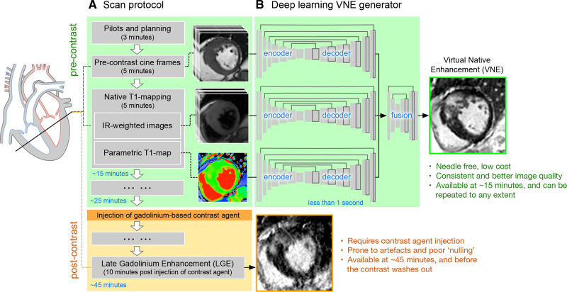
Late gadolinium enhancement (LGE) cardiovascular magnetic resonance (CMR) imaging is the gold standard for noninvasive myocardial tissue characterization but requires intravenous contrast agent administration. It is highly desired to develop a contrast agent-free technology to replace LGE for faster and cheaper CMR scans.

METHODS

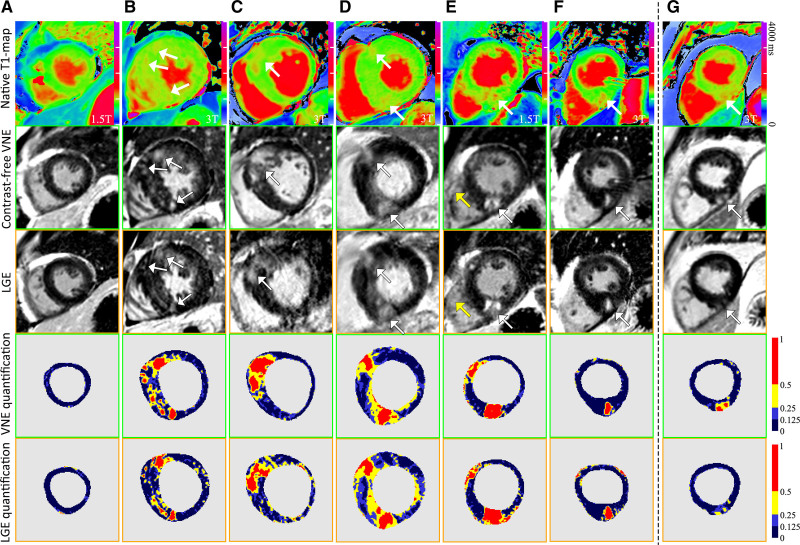
A CMR virtual native enhancement (VNE) imaging technology was developed using artificial intelligence. The deep learning model for generating VNE uses multiple streams of convolutional neural networks to exploit and enhance the existing signals in native T1 maps (pixel-wise maps of tissue T1 relaxation times) and cine imaging of cardiac structure and function, presenting them as LGE-equivalent images. The VNE generator was trained using generative adversarial networks. This technology was first developed on CMR datasets from the multicenter Hypertrophic Cardiomyopathy Registry, using hypertrophic cardiomyopathy as an exemplar. The datasets were randomized into 2 independent groups for deep learning training and testing. The test data of VNE and LGE were scored and contoured by experienced human operators to assess image quality, visuospatial agreement, and myocardial lesion burden quantification. Image quality was compared using a nonparametric Wilcoxon test. Intra- and interobserver agreement was analyzed using intraclass correlation coefficients (ICC). Lesion quantification by VNE and LGE were compared using linear regression and ICC.

RESULTS

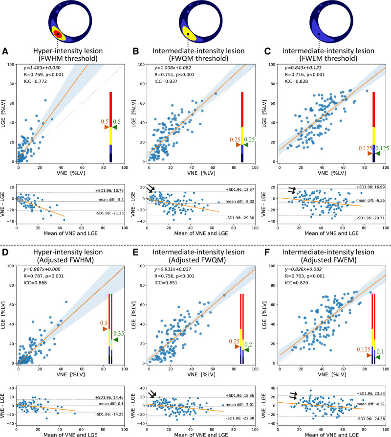
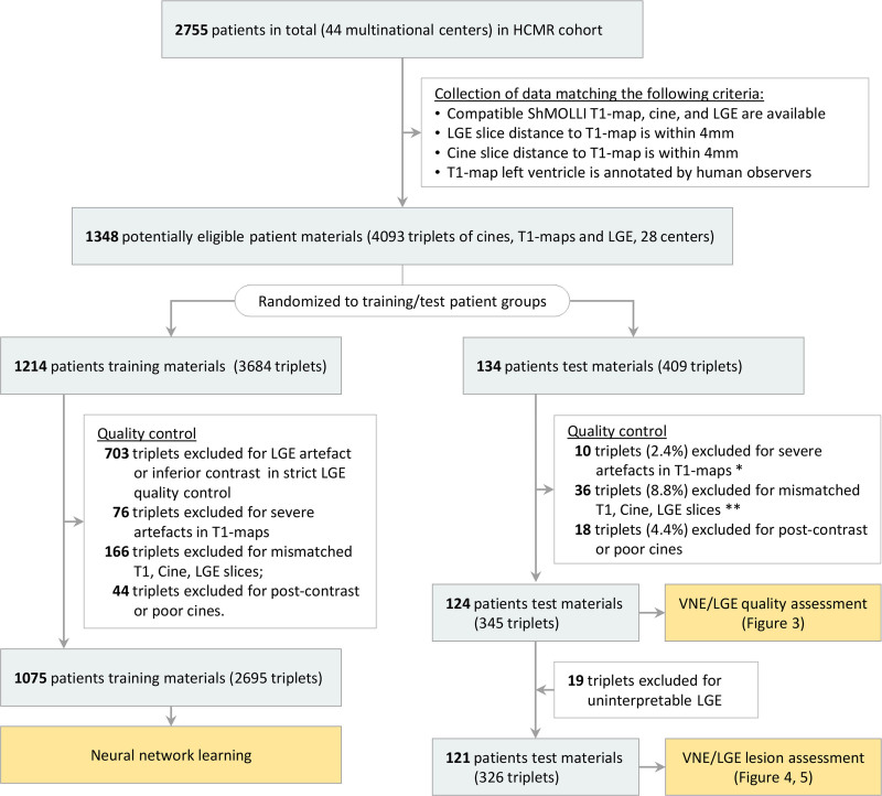
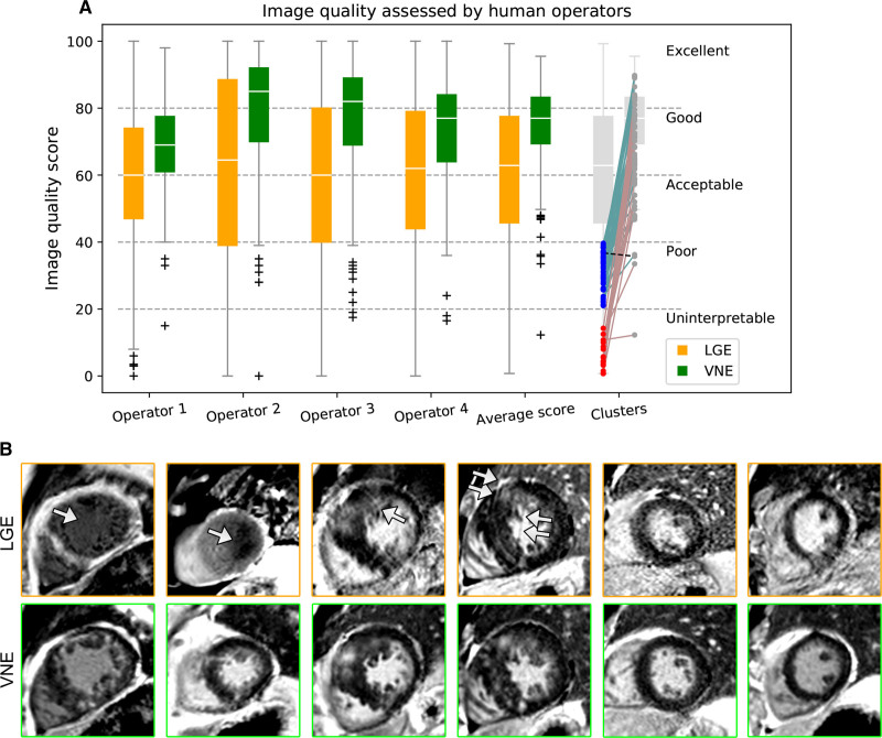
A total of 1348 hypertrophic cardiomyopathy patients provided 4093 triplets of matched T1 maps, cines, and LGE datasets. After randomization and data quality control, 2695 datasets were used for VNE method development and 345 were used for independent testing. VNE had significantly better image quality than LGE, as assessed by 4 operators (n=345 datasets; <0.001 [Wilcoxon test]). VNE revealed lesions characteristic of hypertrophic cardiomyopathy in high visuospatial agreement with LGE. In 121 patients (n=326 datasets), VNE correlated with LGE in detecting and quantifying both hyperintensity myocardial lesions (=0.77-0.79; ICC=0.77-0.87; <0.001) and intermediate-intensity lesions (=0.70-0.76; ICC=0.82-0.85; <0.001). The native CMR images (cine plus T1 map) required for VNE can be acquired within 15 minutes and producing a VNE image takes less than 1 second.

CONCLUSIONS

VNE is a new CMR technology that resembles conventional LGE but without the need for contrast administration. VNE achieved high agreement with LGE in the distribution and quantification of lesions, with significantly better image quality.

摘要

背景

晚期钆增强(LGE)心血管磁共振(CMR)成像技术是无创性心肌组织特征描述的金标准,但需要静脉内造影剂给药。人们非常希望开发一种无造影剂的技术来替代 LGE,以实现更快和更便宜的 CMR 扫描。

方法

使用人工智能开发了一种 CMR 虚拟原生增强(VNE)成像技术。用于生成 VNE 的深度学习模型使用多个卷积神经网络流来利用和增强原生 T1 图谱(组织 T1 弛豫时间的像素图谱)和心脏结构和功能的电影成像中的现有信号,将其呈现为 LGE 等效图像。VNE 生成器使用生成对抗网络进行训练。该技术首先在多中心肥厚型心肌病注册中心的 CMR 数据集上开发,以肥厚型心肌病为例。数据集被随机分为 2 个独立的组进行深度学习训练和测试。VNE 和 LGE 的测试数据由有经验的操作人员进行评分和描绘,以评估图像质量、视觉空间一致性和心肌病变负担定量。使用非参数 Wilcoxon 检验比较图像质量。使用组内相关系数(ICC)分析内部和观察者之间的一致性。使用线性回归和 ICC 比较 VNE 和 LGE 的病变定量。

结果

共有 1348 例肥厚型心肌病患者提供了 4093 对匹配的 T1 图谱、电影和 LGE 数据集。随机化和数据质量控制后,使用 2695 个数据集进行 VNE 方法开发,使用 345 个数据集进行独立测试。VNE 在 4 名操作人员(n=345 个数据集;<0.001[Wilcoxon 检验])评估的图像质量明显优于 LGE。VNE 以高视觉空间一致性显示出与 LGE 一致的肥厚型心肌病特征性病变。在 121 例患者(n=326 个数据集)中,VNE 与 LGE 在检测和定量高信号心肌病变(=0.77-0.79;ICC=0.77-0.87;<0.001)和中等强度病变(=0.70-0.76;ICC=0.82-0.85;<0.001)方面具有相关性。VNE 所需的原生 CMR 图像(电影加 T1 图谱)可在 15 分钟内采集,生成 VNE 图像不到 1 秒。

结论

VNE 是一种新的 CMR 技术,类似于传统的 LGE,但无需造影剂给药。VNE 在病变的分布和定量方面与 LGE 具有高度一致性,并且图像质量显著提高。

https://cdn.ncbi.nlm.nih.gov/pmc/blobs/0155/8378544/02007c6325dd/cir-144-589-g005.jpg
https://cdn.ncbi.nlm.nih.gov/pmc/blobs/0155/8378544/4f63bb9ccaf2/cir-144-589-g001.jpg
https://cdn.ncbi.nlm.nih.gov/pmc/blobs/0155/8378544/60c5ebeb2f3f/cir-144-589-g002.jpg
https://cdn.ncbi.nlm.nih.gov/pmc/blobs/0155/8378544/d0d4eae8c0b9/cir-144-589-g003.jpg
https://cdn.ncbi.nlm.nih.gov/pmc/blobs/0155/8378544/161013eddde5/cir-144-589-g004.jpg
https://cdn.ncbi.nlm.nih.gov/pmc/blobs/0155/8378544/02007c6325dd/cir-144-589-g005.jpg

相似文献

1
Toward Replacing Late Gadolinium Enhancement With Artificial Intelligence Virtual Native Enhancement for Gadolinium-Free Cardiovascular Magnetic Resonance Tissue Characterization in Hypertrophic Cardiomyopathy.用人工智能虚拟原生增强替代晚期钆增强,实现肥厚型心肌病的无钆心血管磁共振组织特征分析。
Circulation. 2021 Aug 24;144(8):589-599. doi: 10.1161/CIRCULATIONAHA.121.054432. Epub 2021 Jul 7.
2
Artificial Intelligence for Contrast-Free MRI: Scar Assessment in Myocardial Infarction Using Deep Learning-Based Virtual Native Enhancement.人工智能在无对比剂 MRI 中的应用:基于深度学习的虚拟自然增强在心肌梗死瘢痕评估中的应用。
Circulation. 2022 Nov 15;146(20):1492-1503. doi: 10.1161/CIRCULATIONAHA.122.060137. Epub 2022 Sep 20.
3
Quality control-driven deep ensemble for accountable automated segmentation of cardiac magnetic resonance LGE and VNE images.用于心脏磁共振延迟强化(LGE)和心室非增强(VNE)图像可问责自动分割的质量控制驱动深度集成方法。
Front Cardiovasc Med. 2023 Sep 11;10:1213290. doi: 10.3389/fcvm.2023.1213290. eCollection 2023.
4
Improved Quantification of Myocardium Scar in Late Gadolinium Enhancement Images: Deep Learning Based Image Fusion Approach.基于深度学习的图像融合方法提高钆延迟增强图像中心肌瘢痕的定量评估。
J Magn Reson Imaging. 2021 Jul;54(1):303-312. doi: 10.1002/jmri.27555. Epub 2021 Feb 17.
5
Native T1-mapping for non-contrast assessment of myocardial fibrosis in patients with hypertrophic cardiomyopathy--comparison with late enhancement quantification.肥厚型心肌病患者心肌纤维化非对比评估的固有T1映射——与延迟强化定量的比较。
Magn Reson Imaging. 2015 Jul;33(6):718-24. doi: 10.1016/j.mri.2015.04.001. Epub 2015 Apr 8.
6
Texture signatures of native myocardial T as novel imaging markers for identification of hypertrophic cardiomyopathy patients without scar.天然心肌T的纹理特征作为识别无瘢痕肥厚型心肌病患者的新型成像标志物。
J Magn Reson Imaging. 2020 Sep;52(3):906-919. doi: 10.1002/jmri.27048. Epub 2020 Jan 23.
7
Evaluation of fully automated myocardial segmentation techniques in native and contrast-enhanced T1-mapping cardiovascular magnetic resonance images using fully convolutional neural networks.使用全卷积神经网络评估 native 和 contrast-enhanced T1-mapping 心血管磁共振成像中的全自动心肌分割技术。
Med Phys. 2021 Jan;48(1):215-226. doi: 10.1002/mp.14574. Epub 2020 Dec 1.
8
Endogenous T1ρ cardiovascular magnetic resonance in hypertrophic cardiomyopathy.肥厚型心肌病中的内源性 T1ρ 心血管磁共振。
J Cardiovasc Magn Reson. 2021 Oct 25;23(1):120. doi: 10.1186/s12968-021-00813-5.
9
Predicting Late Gadolinium Enhancement of Acute Myocardial Infarction in Contrast-Free Cardiac Cine MRI Using Deep Generative Learning.基于深度生成式学习的对比剂-free 心脏电影 MRI 预测急性心肌梗死的延迟钆增强。
Circ Cardiovasc Imaging. 2024 Sep;17(9):e016786. doi: 10.1161/CIRCIMAGING.124.016786. Epub 2024 Sep 10.
10
Automatic machine learning based on native T1 mapping can identify myocardial fibrosis in patients with hypertrophic cardiomyopathy.基于原生 T1 映射的自动机器学习可以识别肥厚型心肌病患者的心肌纤维化。
Eur Radiol. 2022 Feb;32(2):1044-1053. doi: 10.1007/s00330-021-08228-7. Epub 2021 Sep 3.

引用本文的文献

1
Artificial intelligence in medicine: Current applications in cardiology, oncology, and radiology.医学中的人工智能:当前在心脏病学、肿瘤学和放射学中的应用。
World J Methodol. 2025 Dec 20;15(4):106854. doi: 10.5662/wjm.v15.i4.106854.
2
Artificial intelligence for cardiac imaging is ready for widespread clinical use: Pro Con debate AI for cardiac imaging.用于心脏成像的人工智能已准备好广泛应用于临床:支持与反对用于心脏成像的人工智能的辩论
BJR Open. 2025 Jun 6;7(1):tzaf015. doi: 10.1093/bjro/tzaf015. eCollection 2025 Jan.
3
Cardiac Magnetic Resonance in the Evaluation and Management of Nonischemic Cardiomyopathies.

本文引用的文献

1
Cardiac segmentation on late gadolinium enhancement MRI: A benchmark study from multi-sequence cardiac MR segmentation challenge.延迟钆增强磁共振成像的心脏分割:来自多序列心脏磁共振分割挑战赛的一项基准研究。
Med Image Anal. 2022 Oct;81:102528. doi: 10.1016/j.media.2022.102528. Epub 2022 Jul 9.
2
Deep neural network ensemble for on-the-fly quality control-driven segmentation of cardiac MRI T1 mapping.基于深度神经网络集成的心脏 MRI T1 映射实时质量控制驱动分割。
Med Image Anal. 2021 Jul;71:102029. doi: 10.1016/j.media.2021.102029. Epub 2021 Mar 11.
3
Quality assurance of quantitative cardiac T1-mapping in multicenter clinical trials - A T1 phantom program from the hypertrophic cardiomyopathy registry (HCMR) study.
心脏磁共振成像在非缺血性心肌病评估与管理中的应用
JACC Heart Fail. 2025 Sep;13(9):102525. doi: 10.1016/j.jchf.2025.102525. Epub 2025 Jul 5.
4
Toward understanding the balanced steady-state free precession signal intensity changes in cine cardiac magnetic resonance imaging: A preliminary evaluation in healthy subjects pre- and postcontrast.关于理解心脏电影磁共振成像中平衡稳态自由进动信号强度变化:对比剂注射前后健康受试者的初步评估
J Cardiovasc Magn Reson. 2025 May 8;27(2):101908. doi: 10.1016/j.jocmr.2025.101908.
5
Next-Generation Cardiac Magnetic Resonance Imaging Techniques for Characterization of Myocardial Disease.用于心肌疾病特征描述的下一代心脏磁共振成像技术
Curr Treat Options Cardiovasc Med. 2024 Aug;26(8):243-254. doi: 10.1007/s11936-024-01044-4. Epub 2024 Aug 9.
6
Cardiac Magnetic Resonance Guidance for the Pathogenetic Definition of Cardiomyopathies.心肌病发病机制定义的心脏磁共振引导
Curr Cardiol Rep. 2025 Apr 16;27(1):85. doi: 10.1007/s11886-025-02233-8.
7
The role of artificial intelligence in cardiovascular research: Fear less and live bolder.人工智能在心血管研究中的作用:少些恐惧,大胆生活。
Eur J Clin Invest. 2025 Apr;55 Suppl 1(Suppl 1):e14364. doi: 10.1111/eci.14364.
8
Artificial intelligence in medical imaging: From task-specific models to large-scale foundation models.医学影像中的人工智能:从特定任务模型到大规模基础模型。
Chin Med J (Engl). 2025 Mar 20;138(6):651-663. doi: 10.1097/CM9.0000000000003489. Epub 2025 Feb 26.
9
Applications of Artificial Intelligence for the Prediction and Diagnosis of Cancer Therapy-Related Cardiac Dysfunction in Oncology Patients.人工智能在肿瘤患者癌症治疗相关心脏功能障碍预测与诊断中的应用
Cancers (Basel). 2025 Feb 11;17(4):605. doi: 10.3390/cancers17040605.
10
[Clincial Research Progress in Using Magnetic Resonance Imaging to Assess Myocardial Fibrosis in Hypertrophic Cardiomyopathy].[磁共振成像评估肥厚型心肌病心肌纤维化的临床研究进展]
Sichuan Da Xue Xue Bao Yi Xue Ban. 2024 Nov 20;55(6):1357-1363. doi: 10.12182/20241160601.
多中心临床试验中心脏 T1 mapping 定量的质量保证 - 肥厚型心肌病注册研究(HCMR)的 T1 体模项目。
Int J Cardiol. 2021 May 1;330:251-258. doi: 10.1016/j.ijcard.2021.01.026. Epub 2021 Jan 31.
4
Deep learning with attention supervision for automated motion artefact detection in quality control of cardiac T1-mapping.用于心脏T1映射质量控制中自动运动伪影检测的带注意力监督的深度学习
Artif Intell Med. 2020 Nov;110:101955. doi: 10.1016/j.artmed.2020.101955. Epub 2020 Sep 7.
5
2020 AHA/ACC Guideline for the Diagnosis and Treatment of Patients With Hypertrophic Cardiomyopathy: A Report of the American College of Cardiology/American Heart Association Joint Committee on Clinical Practice Guidelines.2020年美国心脏协会/美国心脏病学会肥厚型心肌病患者诊断和治疗指南:美国心脏病学会/美国心脏协会临床实践指南联合委员会报告
Circulation. 2020 Dec 22;142(25):e558-e631. doi: 10.1161/CIR.0000000000000937. Epub 2020 Nov 20.
6
Mavacamten Favorably Impacts Cardiac Structure in Obstructive Hypertrophic Cardiomyopathy: EXPLORER-HCM Cardiac Magnetic Resonance Substudy Analysis.马伐卡坦对梗阻性肥厚型心肌病的心脏结构有积极影响:EXPLORER-HCM心脏磁共振亚研究分析。
Circulation. 2021 Feb 9;143(6):606-608. doi: 10.1161/CIRCULATIONAHA.120.052359. Epub 2020 Nov 15.
7
Standardization of T1-mapping in cardiovascular magnetic resonance using clustered structuring for benchmarking normal ranges.采用聚类结构对心血管磁共振 T1 映射进行标准化,用于基准正常范围。
Int J Cardiol. 2021 Mar 1;326:220-225. doi: 10.1016/j.ijcard.2020.10.041. Epub 2020 Oct 21.
8
Cardiovascular Magnetic Resonance for the Differentiation of Left Ventricular Hypertrophy.心血管磁共振在左心室肥厚鉴别诊断中的应用。
Curr Heart Fail Rep. 2020 Oct;17(5):192-204. doi: 10.1007/s11897-020-00481-z.
9
Standardized cardiovascular magnetic resonance imaging (CMR) protocols: 2020 update.标准化心血管磁共振成像(CMR)协议:2020 年更新。
J Cardiovasc Magn Reson. 2020 Feb 24;22(1):17. doi: 10.1186/s12968-020-00607-1.
10
MRI T1 Mapping in Hypertrophic Cardiomyopathy: Evaluation in Patients Without Late Gadolinium Enhancement and Hemodynamic Obstruction.磁共振 T1 Mapping 在肥厚型心肌病中的应用:无延迟钆增强和血流动力学梗阻患者的评估。
Radiology. 2020 Feb;294(2):275-286. doi: 10.1148/radiol.2019190651. Epub 2019 Nov 26.