Department of Radiology, Cancer Center, Zhejiang Provincial People's Hospital, Affiliated People's Hospital, Hangzhou Medical College, Hangzhou, Zhejiang, China.
BMC Cancer. 2022 Sep 3;22(1):949. doi: 10.1186/s12885-022-10036-1.
To evaluate the difference between multiple primary lung adenocarcinoma (MPLA) and solitary primary lung adenocarcinoma (SPLA) by delta-radiomics based machine learning algorithms in CT images.
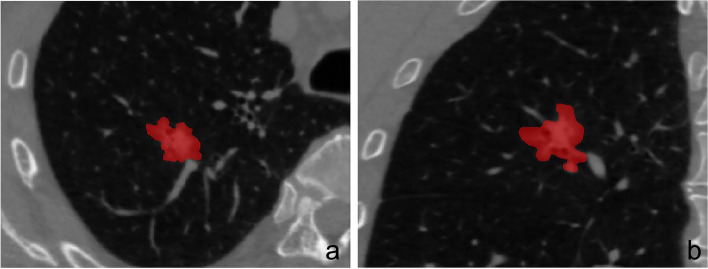
A total of 1094 patients containing 268 MPLAs and 826 SPLAs were recruited for this retrospective study between 2014 to 2020. After the segmentation of volume of interest, the radiomic features were automatically calculated. The patients were categorized into the training set and testing set by a random proportion of 7:3. After feature selection, the relevant classifiers were constructed by the machine learning algorithms of Bayes, forest, k-nearest neighbor, logistic regression, support vector machine, and decision tree. The relative standard deviation (RSD) was calculated and the classification model with minimal RSD was chosen for delta-radiomics analysis to explore the variation of tumor during follow-up surveillance in the cohort of 225 MPLAs and 320 SPLAs. According to the different follow-up duration, it was divided into group A (3-12 months), group B (13-24 months), and group C (25-48 months). Then the corresponding delta-radiomics classifiers were developed to predict MPLAs. The area under the receiver operator characteristic curve (AUC) with 95% confidence interval (CI) was quantified to evaluate the efficiency of the model.
To radiomics analysis, the forest classifier (FC-radio) with the minimal RSD showed the better stability with AUCs of 0.840 (95%CI, 0.810-0.867) and 0.670 (95%CI, 0.611-0.724) in the training and testing set. The AUCs of the forest classifier based on delta-radiomics (FC-delta) were higher than those of FC-radio. In addition, with the extension of follow-up duration, the performance of FC-delta in Group C were the best with AUCs of 0.998 (95%CI, 0.993-1.000) in the training set and 0.853 (95%CI, 0.752-0.940) in the testing set.
The machine-learning approach based on radiomics and delta-radiomics helped to differentiate SPLAs from MPLAs. The FC-delta with a longer follow-up duration could better distinguish between SPLAs and MPLAs.
利用基于 delta-radiomics 的机器学习算法在 CT 图像中评估多发性肺腺癌(MPLA)与单发肺腺癌(SPLA)之间的差异。
本回顾性研究共纳入 2014 年至 2020 年间的 1094 例患者,其中包括 268 例 MPLA 和 826 例 SPLA。在进行感兴趣区的分割后,自动计算放射组学特征。通过 7:3 的随机比例将患者分为训练集和测试集。在特征选择后,使用贝叶斯、森林、k-最近邻、逻辑回归、支持向量机和决策树等机器学习算法构建相关分类器。计算相对标准偏差(RSD),选择 RSD 最小的分类模型进行 delta-radiomics 分析,以探讨在 225 例 MPLA 和 320 例 SPLA 队列中肿瘤在随访监测期间的变化。根据不同的随访时间,将其分为 A 组(3-12 个月)、B 组(13-24 个月)和 C 组(25-48 个月)。然后开发相应的 delta-radiomics 分类器以预测 MPLA。通过 95%置信区间(CI)计算的接收者操作特征曲线(ROC)下面积(AUC)来评估模型的效率。
在放射组学分析中,最小 RSD 的森林分类器(FC-radio)在训练集和测试集的 AUC 分别为 0.840(95%CI,0.810-0.867)和 0.670(95%CI,0.611-0.724),显示出更好的稳定性。基于 delta-radiomics 的森林分类器(FC-delta)的 AUC 高于 FC-radio。此外,随着随访时间的延长,FC-delta 在 C 组中的表现最佳,其在训练集和测试集的 AUC 分别为 0.998(95%CI,0.993-1.000)和 0.853(95%CI,0.752-0.940)。
基于放射组学和 delta-radiomics 的机器学习方法有助于区分 SPLA 和 MPLA。FC-delta 随着随访时间的延长,可以更好地区分 SPLA 和 MPLA。