Ma Yanqing, Ma Weijun, Xu Xiren, Cao Fang
Zhejiang Provincial People's Hospital, Hangzhou, China.
Shaoxing City Keqiao District Hospital of Traditional Chinese Medicine, Shaoxing, China.
Front Oncol. 2020 Jul 16;10:1017. doi: 10.3389/fonc.2020.01017. eCollection 2020.
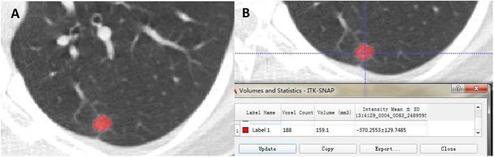
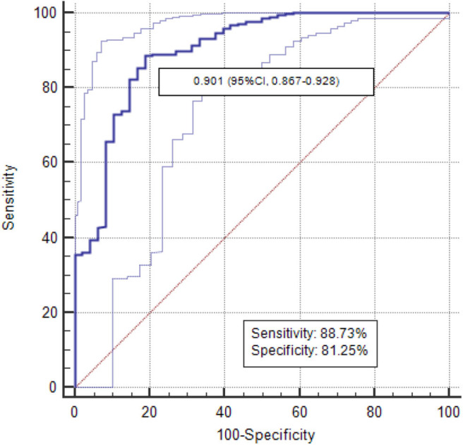
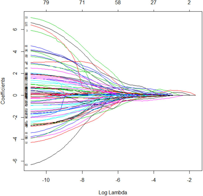
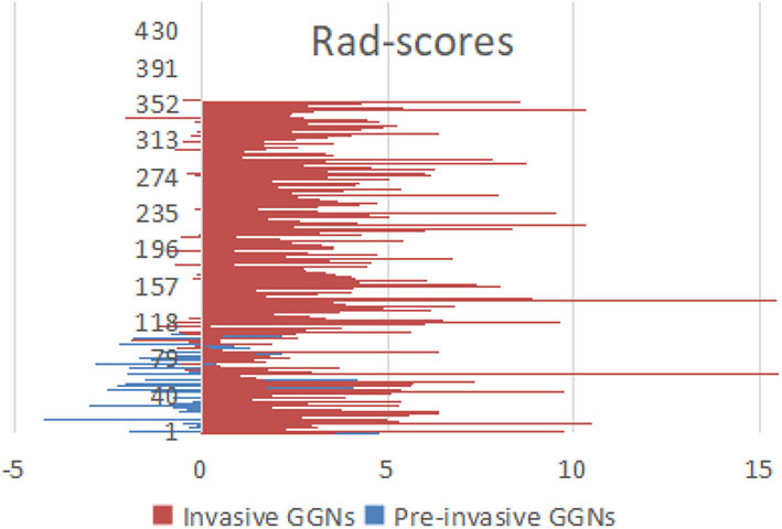
This study aimed to explore the role of delta-radiomics in differentiating pre-invasive ground-glass nodules (GGNs) from invasive GGNs, compared with radiomics signature. A total of 464 patients including 107 pre-invasive GGNs and 357 invasive GGNs were embraced in radiomics signature analysis. 3D regions of interest (ROIs) were contoured with ITK software. By means of ANOVA/MW, correlation analysis, and LASSO, the optimal radiomic features were selected. The logistic classifier of radiomics signature was constructed and radiomic scores (rad-scores) were calculated. A total of 379 patients including 48 pre-invasive GGNs and 331 invasive GGNs with baseline and follow-up CT examinations before surgeries were enrolled in delta-radiomics analysis. Finally, the logistic classifier of delta-radiomics was constructed. The receiver operating characteristic curves (ROCs) were built to evaluate the validity of classifiers. For radiomics signature analysis, six features were selected from 396 radiomic features. The areas under curve (AUCs) of logistic classifiers were 0.865 (95% CI, 0.823-0.900) in the training set and 0.800 (95% CI, 0.724-0.863) in the testing set. The rad-scores of invasive GGNs were larger than those of pre-invasive GGNs. As the follow-up interval went on, more and more delta-radiomic features became statistically different. The AUC of the delta-radiomics logistic classifier was 0.901 (95% CI, 0.867-0.928), which was higher than that of the radiomics signature. The radiomics signature contributes to distinguish pre-invasive and invasive GGNs. The rad-scores of invasive GGNs were larger than those of pre-invasive GGNs. More and more delta-radiomic features appeared to be statistically different as follow-up interval prolonged. Delta-radiomics is superior to radiomics signature in differentiating pre-invasive and invasive GGNs.
本研究旨在探讨与影像组学特征相比,δ-影像组学在鉴别侵袭前磨玻璃结节(GGN)与侵袭性GGN中的作用。影像组学特征分析纳入了464例患者,包括107个侵袭前GGN和357个侵袭性GGN。使用ITK软件勾勒出三维感兴趣区(ROI)。通过方差分析/曼-惠特尼检验、相关性分析和套索法,选择最佳影像组学特征。构建影像组学特征的逻辑分类器并计算影像组学分数(rad分数)。δ-影像组学分析纳入了379例患者,包括48个侵袭前GGN和331个侵袭性GGN,这些患者在手术前有基线和随访CT检查。最后,构建δ-影像组学的逻辑分类器。绘制受试者工作特征曲线(ROC)以评估分类器的有效性。对于影像组学特征分析,从396个影像组学特征中选择了6个特征。逻辑分类器在训练集的曲线下面积(AUC)为0.865(95%CI,0.823-0.900),在测试集为0.800(95%CI,0.724-0.863)。侵袭性GGN的rad分数高于侵袭前GGN。随着随访间隔的延长,越来越多的δ-影像组学特征在统计学上出现差异。δ-影像组学逻辑分类器的AUC为0.901(95%CI,0.867-0.928),高于影像组学特征。影像组学特征有助于鉴别侵袭前和侵袭性GGN。侵袭性GGN的rad分数高于侵袭前GGN。随着随访间隔延长,越来越多的δ-影像组学特征在统计学上出现差异。在鉴别侵袭前和侵袭性GGN方面,δ-影像组学优于影像组学特征。