• 文献检索
  • 文档翻译
  • 深度研究
  • 学术资讯
  • Suppr Zotero 插件Zotero 插件
  • 邀请有礼
  • 套餐&价格
  • 历史记录
应用&插件
Suppr Zotero 插件Zotero 插件浏览器插件Mac 客户端Windows 客户端微信小程序
定价
高级版会员购买积分包购买API积分包
服务
文献检索文档翻译深度研究API 文档MCP 服务
关于我们
关于 Suppr公司介绍联系我们用户协议隐私条款
关注我们

Suppr 超能文献

核心技术专利:CN118964589B侵权必究
粤ICP备2023148730 号-1Suppr @ 2026

文献检索

告别复杂PubMed语法,用中文像聊天一样搜索,搜遍4000万医学文献。AI智能推荐,让科研检索更轻松。

立即免费搜索

文件翻译

保留排版,准确专业,支持PDF/Word/PPT等文件格式,支持 12+语言互译。

免费翻译文档

深度研究

AI帮你快速写综述,25分钟生成高质量综述,智能提取关键信息,辅助科研写作。

立即免费体验

对老年社区居住者大脑中扩大的血管周围间隙进行定量测量,与视网膜微血管参数相关。

Quantitative measurements of enlarged perivascular spaces in the brain are associated with retinal microvascular parameters in older community-dwelling subjects.

作者信息

Ballerini Lucia, McGrory Sarah, Valdés Hernández Maria Del C, Lovreglio Ruggiero, Pellegrini Enrico, MacGillivray Tom, Muñoz Maniega Susana, Henderson Ross, Taylor Adele, Bastin Mark E, Doubal Fergus, Trucco Emanuele, Deary Ian J, Wardlaw Joanna

机构信息

Department of Neuroimaging Sciences, Centre for Clinical Brain Sciences, and VAMPIRE Project, University of Edinburgh, Edinburgh, EH16 4SB, UK.

Dementia Research Institute, University of Edinburgh, Edinburgh, UK.

出版信息

Cereb Circ Cogn Behav. 2020;1:100002. doi: 10.1016/j.cccb.2020.100002.

DOI:10.1016/j.cccb.2020.100002
PMID:33458712
原文链接:https://pmc.ncbi.nlm.nih.gov/articles/PMC7792660/
Abstract

BACKGROUND

Perivascular Spaces (PVS) become increasingly visible with advancing age on brain MRI, yet their relationship to morphological changes in the underlying microvessels remains poorly understood. Retinal and cerebral microvessels share morphological and physiological properties. We compared computationally-derived PVS morphologies with retinal vessel morphologies in older people.

METHODS

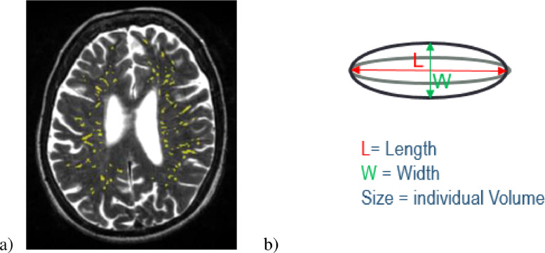
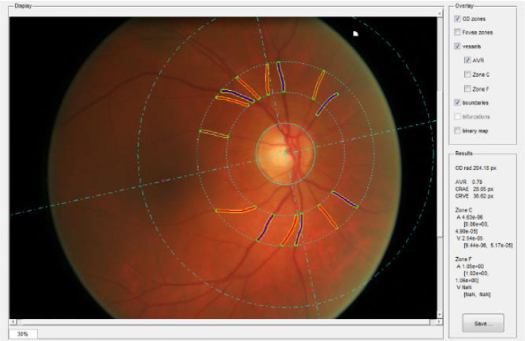
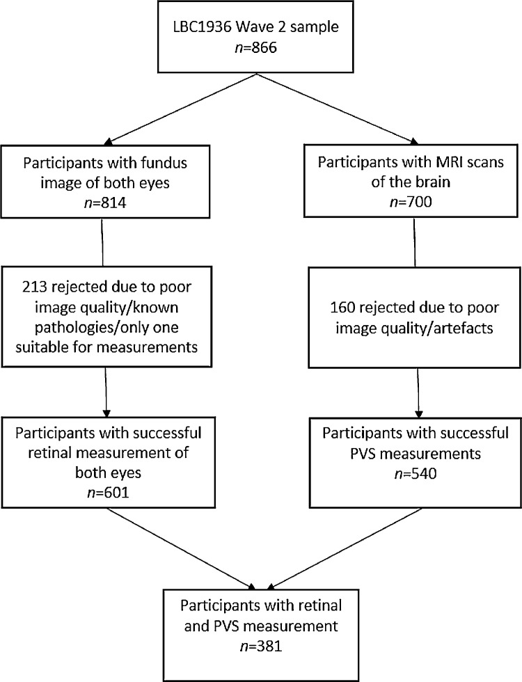
We analysed data from community-dwelling individuals who underwent multimodal brain MRI and retinal fundus camera imaging at mean age 72.55 years (SD=0.71). We assessed centrum semiovale PVS computationally to determine PVS total volume and count, and mean per-subject individual PVS length, width and size. We analysed retinal images using the VAMPIRE software suite, obtaining the Central Retinal Artery and Vein Equivalents (CRVE and CRAE), Arteriole-to-Venule ratio (AVR), and fractal dimension (FD) of both eyes. We investigated associations using general linear models, adjusted for age, gender, and major vascular risk factors.

RESULTS

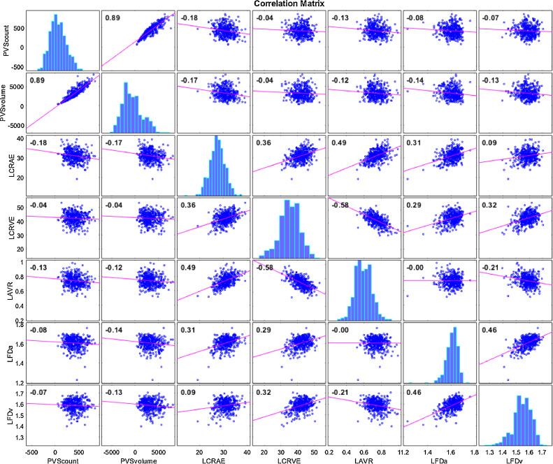
In 381 subjects with all measures, increasing total PVS volume and count were associated with decreased CRAE in the left eye (volume =-0.170, count =-0.184, <0.001). No associations of PVS with CRVE were found. The PVS total volume, individual width and size increased with decreasing FD of the arterioles (a) and venules (v) of the left eye (total volume: FDa =-0.137, FDv =-0.139, <0.01; width: FDa =-0.144, FDv =-0.158, <0.01; size: FDa =-0.157, FDv =-0.162, <0.01).

CONCLUSIONS

Increase in PVS number and size visible on MRI reflect arteriolar narrowing and lower retinal arteriole and venule branching complexity, both markers of impaired microvascular health. Computationally-derived PVS metrics may be an early indicator of failing vascular health and should be tested in longitudinal studies.

摘要

背景

脑磁共振成像(MRI)显示,随着年龄增长,血管周围间隙(PVS)越来越明显,但其与潜在微血管形态变化的关系仍知之甚少。视网膜和脑微血管具有共同的形态和生理特性。我们比较了老年人通过计算得出的PVS形态与视网膜血管形态。

方法

我们分析了社区居住个体的数据,这些个体平均年龄为72.55岁(标准差=0.71),接受了多模态脑MRI和视网膜眼底相机成像。我们通过计算评估半卵圆中心PVS,以确定PVS总体积和数量,以及每个受试者的个体PVS平均长度、宽度和大小。我们使用VAMPIRE软件套件分析视网膜图像,获得双眼的视网膜中央动脉和静脉等效值(CRVE和CRAE)、动静脉比(AVR)和分形维数(FD)。我们使用一般线性模型进行关联研究,并对年龄、性别和主要血管危险因素进行了调整。

结果

在381名接受了所有测量的受试者中,PVS总体积和数量的增加与左眼CRAE的降低相关(体积=-0.170,数量=- 0.184,P<0.001)。未发现PVS与CRVE之间存在关联。左眼小动脉(a)和小静脉(v)的FD降低时,PVS总体积、个体宽度和大小增加(总体积:FDa=-0.137,FDv=-0.139,P<0.01;宽度:FDa=-0.144,FDv=-0.158,P<0.01;大小:FDa=-0.157,FDv=-0.162,P<0.01)。

结论

MRI上可见PVS数量和大小的增加反映了小动脉狭窄以及视网膜小动脉和小静脉分支复杂性降低,这两者都是微血管健康受损的标志。通过计算得出的PVS指标可能是血管健康衰退的早期指标,应在纵向研究中进行检验。

https://cdn.ncbi.nlm.nih.gov/pmc/blobs/9c80/9616227/ef6702b86644/gr4.jpg
https://cdn.ncbi.nlm.nih.gov/pmc/blobs/9c80/9616227/57fdd959978b/gr1.jpg
https://cdn.ncbi.nlm.nih.gov/pmc/blobs/9c80/9616227/0710044039d6/gr2.jpg
https://cdn.ncbi.nlm.nih.gov/pmc/blobs/9c80/9616227/e21647a06dab/gr3.jpg
https://cdn.ncbi.nlm.nih.gov/pmc/blobs/9c80/9616227/ef6702b86644/gr4.jpg
https://cdn.ncbi.nlm.nih.gov/pmc/blobs/9c80/9616227/57fdd959978b/gr1.jpg
https://cdn.ncbi.nlm.nih.gov/pmc/blobs/9c80/9616227/0710044039d6/gr2.jpg
https://cdn.ncbi.nlm.nih.gov/pmc/blobs/9c80/9616227/e21647a06dab/gr3.jpg
https://cdn.ncbi.nlm.nih.gov/pmc/blobs/9c80/9616227/ef6702b86644/gr4.jpg

相似文献

1
Quantitative measurements of enlarged perivascular spaces in the brain are associated with retinal microvascular parameters in older community-dwelling subjects.对老年社区居住者大脑中扩大的血管周围间隙进行定量测量,与视网膜微血管参数相关。
Cereb Circ Cogn Behav. 2020;1:100002. doi: 10.1016/j.cccb.2020.100002.
2
Computational quantification of brain perivascular space morphologies: Associations with vascular risk factors and white matter hyperintensities. A study in the Lothian Birth Cohort 1936.计算脑小血管周围间隙形态学的定量分析:与血管危险因素和脑白质高信号的相关性。在洛锡安出生队列 1936 年的一项研究。
Neuroimage Clin. 2020;25:102120. doi: 10.1016/j.nicl.2019.102120. Epub 2019 Dec 9.
3
Perivascular spaces in the centrum semiovale at the beginning of the 8th decade of life: effect on cognition and associations with mineral deposition.脑白质半卵圆中心第 8 个 10 年开始的血管周围间隙:对认知的影响及其与矿物质沉积的关系。
Brain Imaging Behav. 2020 Oct;14(5):1865-1875. doi: 10.1007/s11682-019-00128-1.
4
Determination of retinal blood vessel diameters and arteriovenous ratios in systemic hypertension: comparison of different calculation formulae.系统性高血压中视网膜血管直径及动静脉比率的测定:不同计算公式的比较
Graefes Arch Clin Exp Ophthalmol. 2007 Jan;245(1):8-17. doi: 10.1007/s00417-006-0358-7. Epub 2006 Jul 11.
5
Sleep quality, perivascular spaces and brain health markers in ageing - A longitudinal study in the Lothian Birth Cohort 1936.衰老过程中的睡眠质量、血管周围间隙与脑健康标志物——对1936年洛锡安出生队列的一项纵向研究
Sleep Med. 2023 Jun;106:123-131. doi: 10.1016/j.sleep.2023.03.016. Epub 2023 Mar 16.
6
Relationship Between Venules and Perivascular Spaces in Sporadic Small Vessel Diseases.散发性小血管病中小静脉与血管周围腔隙的关系。
Stroke. 2020 May;51(5):1503-1506. doi: 10.1161/STROKEAHA.120.029163. Epub 2020 Apr 8.
7
Lower Retinal Arteriolar Density Is Associated With Higher Cerebral Small Vessel Disease Burden: An Optical Coherence Tomography Angiography Study.视网膜小动脉密度降低与更高的脑小血管病负担相关:一项光学相干断层扫描血管造影研究。
Brain Behav. 2025 Feb;15(2):e70342. doi: 10.1002/brb3.70342.
8
Retinal vessel phenotype in patients with primary open-angle glaucoma.原发性开角型青光眼患者的视网膜血管表型。
Acta Ophthalmol. 2020 Feb;98(1):e88-e93. doi: 10.1111/aos.14192. Epub 2019 Jul 29.
9
Retinal vessel caliber among people with acquired immunodeficiency syndrome: relationships with disease-associated factors and mortality.获得性免疫缺陷综合征患者的视网膜血管口径:与疾病相关因素和死亡率的关系。
Am J Ophthalmol. 2012 Mar;153(3):434-444.e1. doi: 10.1016/j.ajo.2011.08.028. Epub 2011 Oct 22.
10
On the quantitative effects of compression of retinal fundus images on morphometric vascular measurements in VAMPIRE.关于视网膜眼底图像压缩对VAMPIRE中形态学血管测量的定量影响
Comput Methods Programs Biomed. 2021 Apr;202:105969. doi: 10.1016/j.cmpb.2021.105969. Epub 2021 Feb 5.

引用本文的文献

1
Lower Retinal Arteriolar Density Is Associated With Higher Cerebral Small Vessel Disease Burden: An Optical Coherence Tomography Angiography Study.视网膜小动脉密度降低与更高的脑小血管病负担相关:一项光学相干断层扫描血管造影研究。
Brain Behav. 2025 Feb;15(2):e70342. doi: 10.1002/brb3.70342.
2
Perivascular space imaging during therapy for medulloblastoma.髓母细胞瘤治疗期间的血管周围间隙成像
PLoS One. 2025 Feb 7;20(2):e0318278. doi: 10.1371/journal.pone.0318278. eCollection 2025.
3
Editorial. : The journal at age 5.社论:该期刊创刊五周年

本文引用的文献

1
Computational quantification of brain perivascular space morphologies: Associations with vascular risk factors and white matter hyperintensities. A study in the Lothian Birth Cohort 1936.计算脑小血管周围间隙形态学的定量分析:与血管危险因素和脑白质高信号的相关性。在洛锡安出生队列 1936 年的一项研究。
Neuroimage Clin. 2020;25:102120. doi: 10.1016/j.nicl.2019.102120. Epub 2019 Dec 9.
2
Evaluation of the 3D fractal dimension as a marker of structural brain complexity in multiple-acquisition MRI.评估三维分形维数作为多采集 MRI 中结构脑复杂性的标志物。
Hum Brain Mapp. 2019 Aug 1;40(11):3299-3320. doi: 10.1002/hbm.24599. Epub 2019 May 15.
3
Cereb Circ Cogn Behav. 2024 Sep 7;7:100368. doi: 10.1016/j.cccb.2024.100368. eCollection 2024.
4
Association of optic disc pallor and RNFL thickness with cerebral small vessel disease in the PREVENT-Dementia study.在预防痴呆研究中视盘苍白和视网膜神经纤维层厚度与脑小血管病的关联
Alzheimers Dement (Amst). 2024 Aug 8;16(3):e12633. doi: 10.1002/dad2.12633. eCollection 2024 Jul-Sep.
5
A narrative review of retinal vascular parameters and the applications (Part II): Diagnosis in stroke.视网膜血管参数及其应用的叙述性综述(第二部分):中风诊断
Brain Circ. 2023 Sep 27;9(3):129-134. doi: 10.4103/bc.bc_9_23. eCollection 2023 Jul-Sep.
6
A critical guide to the automated quantification of perivascular spaces in magnetic resonance imaging.磁共振成像中血管周围间隙自动定量的关键指南
Front Neurosci. 2022 Dec 14;16:1021311. doi: 10.3389/fnins.2022.1021311. eCollection 2022.
7
Retinal capillary microvessel morphology changes are associated with vascular damage and dysfunction in cerebral small vessel disease.视网膜毛细血管微血管形态的变化与脑小血管病中的血管损伤和功能障碍有关。
J Cereb Blood Flow Metab. 2023 Feb;43(2):231-240. doi: 10.1177/0271678X221135658. Epub 2022 Oct 27.
8
Retinal microvasculature and imaging markers of brain frailty in normal aging adults.正常衰老成年人视网膜微血管与脑衰弱的影像标志物
Front Aging Neurosci. 2022 Aug 22;14:945964. doi: 10.3389/fnagi.2022.945964. eCollection 2022.
9
Characterization of perivascular space pathology in a rat model of cerebral small vessel disease by magnetic resonance imaging.利用磁共振成像对脑小血管病大鼠模型的血管周围间隙病理进行特征描述。
J Cereb Blood Flow Metab. 2022 Oct;42(10):1813-1826. doi: 10.1177/0271678X221105668. Epub 2022 Jun 8.
Retinal microvasculature and cerebral small vessel disease in the Lothian Birth Cohort 1936 and Mild Stroke Study.
洛锡安出生队列 1936 研究和轻度中风研究中的视网膜微血管和脑小血管疾病。
Sci Rep. 2019 Apr 19;9(1):6320. doi: 10.1038/s41598-019-42534-x.
4
Perivascular spaces and their associations with risk factors, clinical disorders and neuroimaging features: A systematic review and meta-analysis.血管周围间隙及其与危险因素、临床疾病和神经影像学特征的关系:系统评价和荟萃分析。
Int J Stroke. 2019 Jun;14(4):359-371. doi: 10.1177/1747493019830321. Epub 2019 Feb 14.
5
Clinical Significance of Magnetic Resonance Imaging Markers of Vascular Brain Injury: A Systematic Review and Meta-analysis.磁共振血管损伤标志物的临床意义:系统评价和荟萃分析。
JAMA Neurol. 2019 Jan 1;76(1):81-94. doi: 10.1001/jamaneurol.2018.3122.
6
Enlarged perivascular spaces in brain MRI: Automated quantification in four regions.脑 MRI 中的血管周围间隙扩大:四个区域的自动量化。
Neuroimage. 2019 Jan 15;185:534-544. doi: 10.1016/j.neuroimage.2018.10.026. Epub 2018 Oct 13.
7
Understanding the role of the perivascular space in cerebral small vessel disease.了解血管周围间隙在脑小血管疾病中的作用。
Cardiovasc Res. 2018 Sep 1;114(11):1462-1473. doi: 10.1093/cvr/cvy113.
8
Towards Standardization of Quantitative Retinal Vascular Parameters: Comparison of SIVA and VAMPIRE Measurements in the Lothian Birth Cohort 1936.迈向视网膜血管定量参数的标准化:1936年洛锡安出生队列中SIVA和VAMPIRE测量方法的比较
Transl Vis Sci Technol. 2018 Mar 23;7(2):12. doi: 10.1167/tvst.7.2.12. eCollection 2018 Mar.
9
Retinal Microvascular Abnormalities as Surrogate Markers of Cerebrovascular Ischemic Disease: A Meta-Analysis.视网膜微血管异常作为脑血管缺血性疾病的替代标志物:一项荟萃分析。
J Stroke Cerebrovasc Dis. 2018 Jul;27(7):1960-1968. doi: 10.1016/j.jstrokecerebrovasdis.2018.02.041. Epub 2018 Mar 20.
10
Cohort Profile Update: The Lothian Birth Cohorts of 1921 and 1936.队列简介更新:1921年和1936年的洛锡安出生队列。
Int J Epidemiol. 2018 Aug 1;47(4):1042-1042r. doi: 10.1093/ije/dyy022.