• 文献检索
  • 文档翻译
  • 深度研究
  • 学术资讯
  • Suppr Zotero 插件Zotero 插件
  • 邀请有礼
  • 套餐&价格
  • 历史记录
应用&插件
Suppr Zotero 插件Zotero 插件浏览器插件Mac 客户端Windows 客户端微信小程序
定价
高级版会员购买积分包购买API积分包
服务
文献检索文档翻译深度研究API 文档MCP 服务
关于我们
关于 Suppr公司介绍联系我们用户协议隐私条款
关注我们

Suppr 超能文献

核心技术专利:CN118964589B侵权必究
粤ICP备2023148730 号-1Suppr @ 2026

文献检索

告别复杂PubMed语法,用中文像聊天一样搜索,搜遍4000万医学文献。AI智能推荐,让科研检索更轻松。

立即免费搜索

文件翻译

保留排版,准确专业,支持PDF/Word/PPT等文件格式,支持 12+语言互译。

免费翻译文档

深度研究

AI帮你快速写综述,25分钟生成高质量综述,智能提取关键信息,辅助科研写作。

立即免费体验

单细胞 RNA 测序证实与早期发病的视网膜变性相关的视网膜小胶质细胞激活。

Single cell RNA sequencing confirms retinal microglia activation associated with early onset retinal degeneration.

机构信息

Shiley Eye Institute, University of California, San Diego, 9415 Campus Point Drive, La Jolla, CA, 92093, USA.

Department of Biochemistry, Faculty of Medicine, UNAM, Mexico City, Mexico.

出版信息

Sci Rep. 2022 Sep 10;12(1):15273. doi: 10.1038/s41598-022-19351-w.

DOI:10.1038/s41598-022-19351-w
PMID:36088481
原文链接:https://pmc.ncbi.nlm.nih.gov/articles/PMC9464204/
Abstract

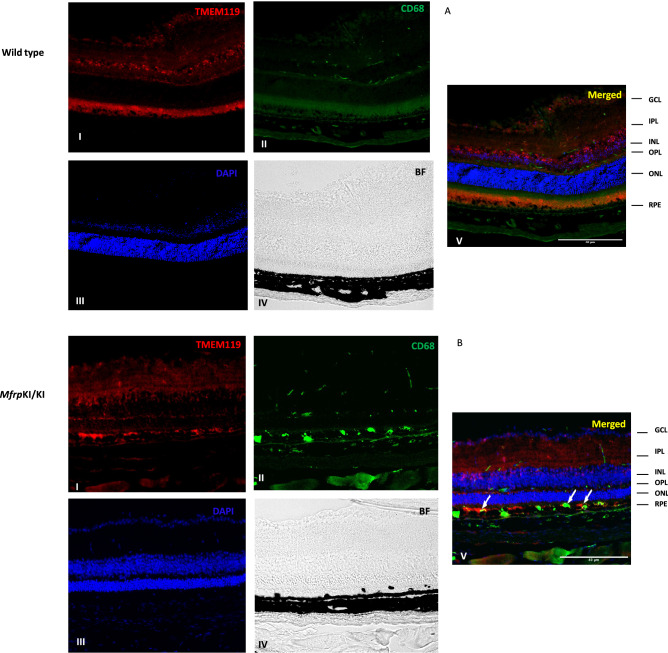
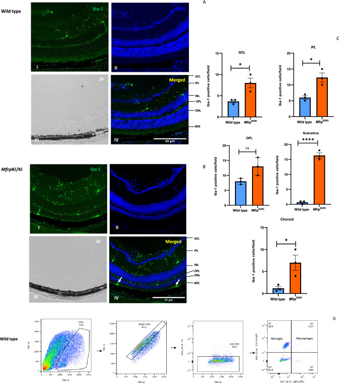
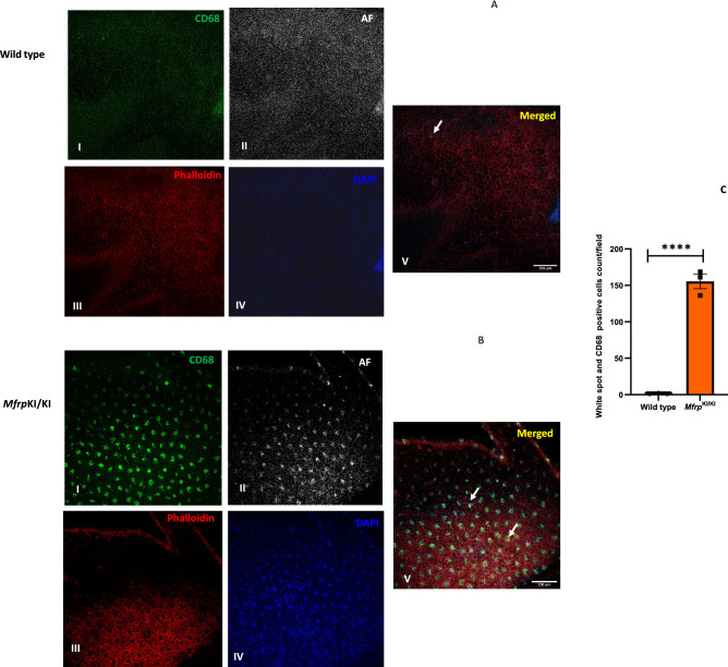
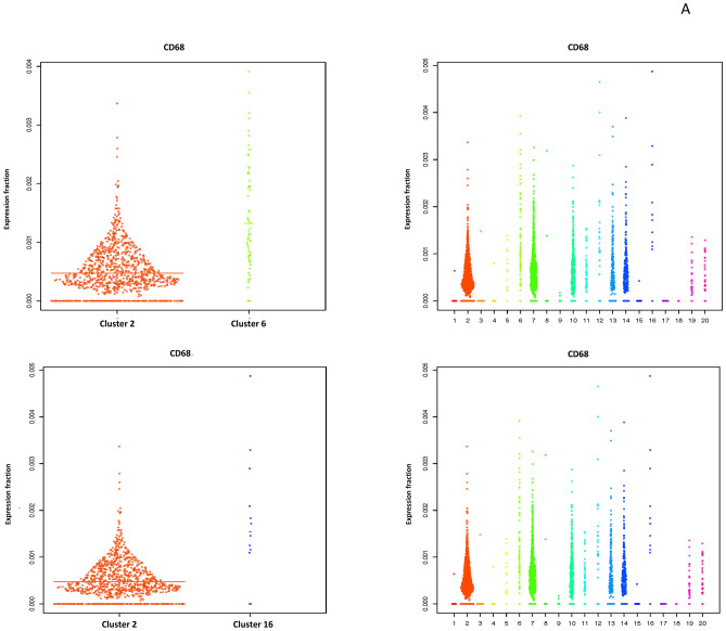
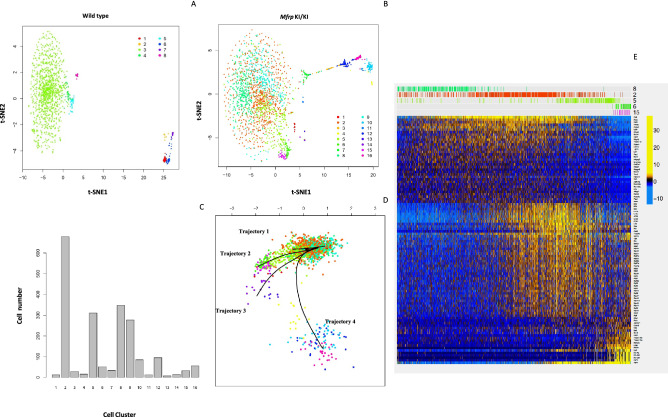
Mutations in the Membrane-type frizzled related protein (Mfrp) gene results in an early-onset retinal degeneration associated with retinitis pigmentosa, microphthalmia, optic disc drusen and foveal schisis. In the current study, a previously characterized mouse model of human retinal degeneration carrying homozygous c.498_499insC mutations in Mfrp (Mfrp) was used. Patients carrying this mutation have retinal degeneration at an early age. The model demonstrates subretinal deposits and develops early-onset photoreceptor degeneration. We observed large subretinal deposits in Mfrp mice which were strongly CD68 positive and co-localized with autofluorescent spots. Single cell RNA sequencing of Mfrp mice retinal microglia showed a significantly higher number of pan-macrophage marker Iba-1 and F4/80 positive cells with increased expression of activation marker (CD68) and lowered microglial homeostatic markers (TMEM119, P2ry13, P2ry13, Siglech) compared with wild type mice confirming microglial activation as observed in retinal immunostaining showing microglia activation in subretinal region. Trajectory analysis identified a small cluster of microglial cells with activation transcriptomic signatures that could represent a subretinal microglia population in Mfrp mice expressing higher levels of APOE. We validated these findings using immunofluorescence staining of retinal cryosections and found a significantly higher number of subretinal Iba-1/ApoE positive microglia in Mfrp mice with some subretinal microglia also expressing lowered levels of microglial homeostatic marker TMEM119, confirming microglial origin. In summary, we confirm that Mfrp mice carrying the c.498_499insC mutation had a significantly higher population of activated microglia in their retina with distinct subsets of subretinal microglia. Further, studies are required to confirm whether the association of increased subretinal microglia in MfrpKI/KI mice are causal in degeneration.

摘要

Mfrp 基因中的突变导致早发性视网膜变性,伴有视网膜色素变性、小眼球、视盘玻璃疣和黄斑裂孔。在本研究中,使用了一种先前描述的携带 Mfrp 基因(Mfrp)中 c.498_499insC 突变的同源纯合子的人类视网膜变性小鼠模型。携带该突变的患者在早期就会出现视网膜变性。该模型表现出视网膜下沉积物,并出现早发性光感受器变性。我们观察到 Mfrp 小鼠中存在大量的视网膜下沉积物,这些沉积物强烈表达 CD68,并与自发荧光斑点共定位。Mfrp 小鼠视网膜小胶质细胞的单细胞 RNA 测序显示,与野生型小鼠相比,具有更多的泛巨噬细胞标志物 Iba-1 和 F4/80 阳性细胞,并且激活标志物(CD68)表达增加,小胶质细胞稳态标志物(TMEM119、P2ry13、P2ry13、Siglech)表达降低,证实了视网膜免疫染色中观察到的小胶质细胞激活,表明小胶质细胞在视网膜下区域激活。轨迹分析确定了一小群具有激活转录组特征的小胶质细胞,这些细胞可能代表 Mfrp 小鼠中表达更高水平 APOE 的视网膜下小胶质细胞群体。我们使用视网膜冷冻切片的免疫荧光染色验证了这些发现,并发现 Mfrp 小鼠中存在更多的视网膜下 Iba-1/ApoE 阳性小胶质细胞,其中一些视网膜下小胶质细胞也表达降低的小胶质细胞稳态标志物 TMEM119,证实了小胶质细胞的起源。总之,我们证实携带 c.498_499insC 突变的 Mfrp 小鼠的视网膜中有明显更多的激活小胶质细胞,并且其视网膜下小胶质细胞存在不同的亚群。还需要进一步的研究来确认 MfrpKI/KI 小鼠中增加的视网膜下小胶质细胞是否与变性有关。

https://cdn.ncbi.nlm.nih.gov/pmc/blobs/f44a/9464204/d42c61673f37/41598_2022_19351_Fig9_HTML.jpg
https://cdn.ncbi.nlm.nih.gov/pmc/blobs/f44a/9464204/d6d8bd7426c3/41598_2022_19351_Fig1_HTML.jpg
https://cdn.ncbi.nlm.nih.gov/pmc/blobs/f44a/9464204/31cd7d47f6ae/41598_2022_19351_Fig2a_HTML.jpg
https://cdn.ncbi.nlm.nih.gov/pmc/blobs/f44a/9464204/223b96cafe4b/41598_2022_19351_Fig3_HTML.jpg
https://cdn.ncbi.nlm.nih.gov/pmc/blobs/f44a/9464204/6cd56a686ca4/41598_2022_19351_Fig4_HTML.jpg
https://cdn.ncbi.nlm.nih.gov/pmc/blobs/f44a/9464204/33f868e4ebc0/41598_2022_19351_Fig5a_HTML.jpg
https://cdn.ncbi.nlm.nih.gov/pmc/blobs/f44a/9464204/8e85b5e8d139/41598_2022_19351_Fig6a_HTML.jpg
https://cdn.ncbi.nlm.nih.gov/pmc/blobs/f44a/9464204/8e17d1625a33/41598_2022_19351_Fig7_HTML.jpg
https://cdn.ncbi.nlm.nih.gov/pmc/blobs/f44a/9464204/966c2d820e14/41598_2022_19351_Fig8_HTML.jpg
https://cdn.ncbi.nlm.nih.gov/pmc/blobs/f44a/9464204/d42c61673f37/41598_2022_19351_Fig9_HTML.jpg

相似文献

1
Single cell RNA sequencing confirms retinal microglia activation associated with early onset retinal degeneration.单细胞 RNA 测序证实与早期发病的视网膜变性相关的视网膜小胶质细胞激活。
Sci Rep. 2022 Sep 10;12(1):15273. doi: 10.1038/s41598-022-19351-w.
2
Long-Term Effects of Gene Therapy in a Novel Mouse Model of Human -Associated Retinopathy.新型人类相关性视网膜病变小鼠模型中的基因治疗的长期效果。
Hum Gene Ther. 2019 May;30(5):632-650. doi: 10.1089/hum.2018.192. Epub 2019 Jan 16.
3
MFRP in Early Onset Retinal Degeneration: Clinical and Molecular Perspectives.早发性视网膜变性中的MFRP:临床与分子视角
Adv Exp Med Biol. 2025;1468:75-79. doi: 10.1007/978-3-031-76550-6_13.
4
A new autosomal recessive syndrome consisting of posterior microphthalmos, retinitis pigmentosa, foveoschisis, and optic disc drusen is caused by a MFRP gene mutation.一种由MFRP基因突变引起的新的常染色体隐性综合征,其特征包括小眼球后段、色素性视网膜炎、黄斑劈裂和视盘玻璃膜疣。
Mol Vis. 2006 Dec 4;12:1483-9.
5
Microglia Inhibition Delays Retinal Degeneration Due to MerTK Phagocytosis Receptor Deficiency.小胶质细胞抑制因 MerTK 吞噬受体缺陷导致的视网膜变性。
Front Immunol. 2020 Jul 16;11:1463. doi: 10.3389/fimmu.2020.01463. eCollection 2020.
6
Expression pattern of Ccr2 and Cx3cr1 in inherited retinal degeneration.Ccr2和Cx3cr1在遗传性视网膜变性中的表达模式
J Neuroinflammation. 2015 Oct 12;12:188. doi: 10.1186/s12974-015-0408-3.
7
Loss of Zebrafish Mfrp Causes Nanophthalmia, Hyperopia, and Accumulation of Subretinal Macrophages.斑马鱼Mfrp缺失导致小眼球、远视以及视网膜下巨噬细胞积聚。
Invest Ophthalmol Vis Sci. 2016 Dec 1;57(15):6805-6814. doi: 10.1167/iovs.16-19593.
8
Mfrp, a gene encoding a frizzled related protein, is mutated in the mouse retinal degeneration 6.Mfrp是一种编码卷曲相关蛋白的基因,在小鼠视网膜变性6中发生突变。
Hum Mol Genet. 2002 Aug 1;11(16):1879-86. doi: 10.1093/hmg/11.16.1879.
9
Differences in the distribution, phenotype and gene expression of subretinal microglia/macrophages in C57BL/6N (Crb1 rd8/rd8) versus C57BL6/J (Crb1 wt/wt) mice.C57BL/6N(Crb1 rd8/rd8)与C57BL6/J(Crb1 wt/wt)小鼠视网膜下小胶质细胞/巨噬细胞在分布、表型和基因表达上的差异。
J Neuroinflammation. 2015 Jan 15;12:6. doi: 10.1186/s12974-014-0221-4.
10
CCL3 production by microglial cells modulates disease severity in murine models of retinal degeneration.小胶质细胞产生的 CCL3 调节视网膜变性的小鼠模型中的疾病严重程度。
J Immunol. 2014 Apr 15;192(8):3816-27. doi: 10.4049/jimmunol.1301738. Epub 2014 Mar 17.

引用本文的文献

1
Spatial transcriptomics reveals regionally altered gene expression that drives retinal degeneration.空间转录组学揭示了驱动视网膜变性的区域基因表达变化。
Commun Biol. 2025 Apr 18;8(1):629. doi: 10.1038/s42003-025-07887-2.
2
Female sex hormones exacerbate retinal neurodegeneration.女性性激素会加剧视网膜神经变性。
Sci Adv. 2025 Apr 11;11(15):eadr6211. doi: 10.1126/sciadv.adr6211.
3
A network medicine approach to investigating ME/CFS pathogenesis in severely ill patients: a pilot study.一种用于研究重症肌痛/慢性疲劳综合征(ME/CFS)患者发病机制的网络医学方法:一项初步研究。

本文引用的文献

1
Circulating inflammatory monocytes oppose microglia and contribute to cone cell death in retinitis pigmentosa.循环炎症单核细胞对抗小胶质细胞并促成色素性视网膜炎中的视锥细胞死亡。
PNAS Nexus. 2022 Mar;1(1). doi: 10.1093/pnasnexus/pgac003. Epub 2022 Mar 2.
2
Cholesterol homeostasis in the vertebrate retina: biology and pathobiology.脊椎动物视网膜中的胆固醇稳态:生物学与病理生物学
J Lipid Res. 2021;62:100057. doi: 10.1194/jlr.TR120000979. Epub 2021 Mar 2.
3
Microglia dynamics in retinitis pigmentosa model: formation of fundus whitening and autofluorescence as an indicator of activity of retinal degeneration.
Front Hum Neurosci. 2025 Feb 10;19:1509346. doi: 10.3389/fnhum.2025.1509346. eCollection 2025.
4
Astragalus polysaccharide attenuates retinal ischemia reperfusion-induced microglial activation through sortilin-related vacuolar protein sorting 10 domain containing receptor 2/laminin subunit alpha 1 upregulation.黄芪多糖通过上调含sortilin相关囊泡蛋白分选10结构域受体2/层粘连蛋白α1亚基减轻视网膜缺血再灌注诱导的小胶质细胞激活。
Cytojournal. 2025 Jan 7;22:2. doi: 10.25259/Cytojournal_131_2024. eCollection 2025.
5
Single Cell Isolation from Human Diabetic Fibrovascular Membranes for Single-Cell RNA Sequencing.从人糖尿病纤维血管膜中分离单细胞用于单细胞RNA测序
Bio Protoc. 2024 Oct 20;14(20):e5096. doi: 10.21769/BioProtoc.5096.
6
Identification of ribosome biogenesis genes and subgroups in ischaemic stroke.鉴定缺血性脑卒中核糖体生物发生基因和亚群。
Front Immunol. 2024 Sep 2;15:1449158. doi: 10.3389/fimmu.2024.1449158. eCollection 2024.
7
Combination of blockade of endothelin signalling and compensation of IGF1 expression protects the retina from degeneration.内皮素信号阻断和 IGF1 表达补偿联合保护视网膜免受变性。
Cell Mol Life Sci. 2024 Jan 22;81(1):51. doi: 10.1007/s00018-023-05087-x.
8
Structure, Function, and Molecular Landscapes of the Aging Retina.衰老视网膜的结构、功能和分子特征。
Annu Rev Vis Sci. 2023 Sep 15;9:177-199. doi: 10.1146/annurev-vision-112122-020950. Epub 2023 May 17.
9
Single-cell transcriptome reveals diversity of Müller cells with different metabolic-mitochondrial signatures in normal and degenerated macula.单细胞转录组揭示了正常和退化黄斑中具有不同代谢-线粒体特征的米勒细胞的多样性。
Front Neurosci. 2022 Dec 22;16:1079498. doi: 10.3389/fnins.2022.1079498. eCollection 2022.
视网膜色素变性模型中的小胶质细胞动力学:眼底白化和自发荧光的形成作为视网膜变性活性的指标。
Sci Rep. 2020 Sep 7;10(1):14700. doi: 10.1038/s41598-020-71626-2.
4
The TSPO-NOX1 axis controls phagocyte-triggered pathological angiogenesis in the eye.TSPO-NOX1 轴控制吞噬细胞触发的眼部病理性血管生成。
Nat Commun. 2020 Jun 1;11(1):2709. doi: 10.1038/s41467-020-16400-8.
5
Microglia versus Monocytes: Distinct Roles in Degenerative Diseases of the Retina.小胶质细胞与单核细胞:在视网膜退行性疾病中的不同作用。
Trends Neurosci. 2020 Jun;43(6):433-449. doi: 10.1016/j.tins.2020.03.012. Epub 2020 Apr 17.
6
Single Eye mRNA-Seq Reveals Normalisation of the Retinal Microglial Transcriptome Following Acute Inflammation.单眼 mRNA-Seq 揭示急性炎症后视网膜小胶质细胞转录组的正常化。
Front Immunol. 2020 Jan 9;10:3033. doi: 10.3389/fimmu.2019.03033. eCollection 2019.
7
CD36 Deficiency Inhibits Retinal Inflammation and Retinal Degeneration in Knockout Mice.CD36 缺乏抑制 基因敲除小鼠的视网膜炎症和变性。
Front Immunol. 2020 Jan 8;10:3032. doi: 10.3389/fimmu.2019.03032. eCollection 2019.
8
Identification of a Unique Subretinal Microglia Type in Retinal Degeneration Using Single Cell RNA-Seq.单细胞 RNA 测序鉴定视网膜变性中小胶质细胞的独特亚型。
Adv Exp Med Biol. 2019;1185:181-186. doi: 10.1007/978-3-030-27378-1_30.
9
Isolation of Region-specific Microglia from One Adult Mouse Brain Hemisphere for Deep Single-cell RNA Sequencing.从一只成年小鼠脑半球中分离区域特异性小胶质细胞用于深度单细胞RNA测序。
J Vis Exp. 2019 Dec 3(154). doi: 10.3791/60347.
10
Annexin A5 regulates surface αvβ5 integrin for retinal clearance phagocytosis.膜联蛋白 A5 调控视网膜清除吞噬作用中的表面 αvβ5 整联蛋白。
J Cell Sci. 2019 Oct 16;132(20):jcs232439. doi: 10.1242/jcs.232439.