Jie Xiao-Xiang, Zhang Meng, Du Ming, Cai Qing-Qing, Cong Qing, Xu Cong-Jian, Zhang Xiao-Yan
Obstetrics and Gynecology Hospital, Fudan University, Shanghai, China.
Shanghai Key Laboratory of Female Reproductive Endocrine Related Diseases, Shanghai, China.
Transl Cancer Res. 2022 Aug;11(8):2636-2646. doi: 10.21037/tcr-22-529.
Circulating tumor cells (CTCs) have considered to be promising liquid biopsy in cancer due to the intact information of whole cells and the potential to reflect micrometastasis. However, CTCs research are extremely limited in ovarian cancer, probably due to their rarity. The predictive value of CTCs and circulating tumor microemboli (CTM) in metastasis remains to be elucidated in ovarian cancer. This study tried to identify CTCs/CTM in ovarian cancer with considerably positive rate. To preliminarily identify the invasive capacity of CTCs/CTM, the epithelial-mesenchymal transition (EMT) patterns of CTCs/CTM was evaluated. Moreover, for comprehensive understanding of invasiveness of disseminated cells in ovarian cancer, EMT pattern of exfoliated tumor cells in ascites were also confirmed in this study.
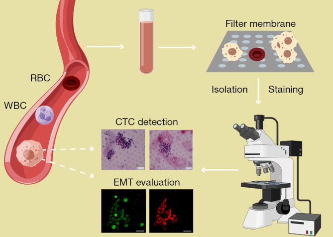
Peripheral blood samples and ascites samples were collected from 22 ovarian cancer patients. The Microfiltration combined with morphological analysis was used to detect CTC single cells or cell clusters. Microfiltration combined with morphological analysis was applied in the CTC isolation and identification. EMT was evaluated by immunofluorescence via markers including vimentin and cytokeratin.
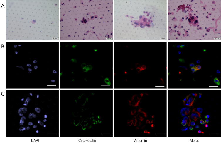
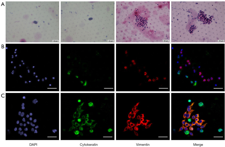
Microfiltration combined with morphological analysis was introduced to detect CTCs/CTM with a positivity rate of 40.9% in ovarian cancer patients. The number of CTC varied from 1 to 8, with CTM number from 4 to 30. CTCs/CTM of all samples have experienced EMT process. Vimentin was expressed in all CTC samples and all tumor cells in ascites, while cytokeratin was expressed in 44.4% (4/9) of CTC samples. There were no significant differences of the clinical parameters between the CTC-positive and CTC-negative patients.
This study showed that both CTCs/CTM and detached tumor cells in ascites might have undergone complete or partial EMT in ovarian cancer. Moreover, microfiltration combined with cytomorphological analysis showed a considerable CTC detection rate.
循环肿瘤细胞(CTCs)因其完整的全细胞信息以及反映微转移的潜力,被认为是癌症中很有前景的液体活检方法。然而,CTCs在卵巢癌中的研究极为有限,可能是由于其稀有性。CTCs和循环肿瘤微栓子(CTM)在卵巢癌转移中的预测价值仍有待阐明。本研究试图在卵巢癌中以相当高的阳性率识别CTCs/CTM。为初步鉴定CTCs/CTM的侵袭能力,评估了CTCs/CTM的上皮-间质转化(EMT)模式。此外,为全面了解卵巢癌中播散细胞的侵袭性,本研究还证实了腹水中脱落肿瘤细胞的EMT模式。
收集22例卵巢癌患者的外周血样本和腹水样本。采用微滤结合形态学分析检测CTCs单细胞或细胞团簇。微滤结合形态学分析应用于CTCs的分离和鉴定。通过免疫荧光法,利用波形蛋白和细胞角蛋白等标志物评估EMT。
引入微滤结合形态学分析检测卵巢癌患者的CTCs/CTM,阳性率为40.9%。CTCs数量从1到8不等,CTM数量从4到30不等。所有样本的CTCs/CTM都经历了EMT过程。波形蛋白在所有CTCs样本和腹水中的所有肿瘤细胞中均有表达,而细胞角蛋白在44.4%(4/9)的CTCs样本中有表达。CTCs阳性和阴性患者的临床参数无显著差异。
本研究表明,卵巢癌中的CTCs/CTM和腹水中的脱落肿瘤细胞可能都经历了完全或部分EMT。此外,微滤结合细胞形态学分析显示CTCs检测率相当可观。