Department of Cell Biology, Yale University School of Medicine, New Haven, CT 06520.
Department of Chemical Engineering, Columbia University, New York, NY 10027.
Proc Natl Acad Sci U S A. 2022 Sep 20;119(38):e2208337119. doi: 10.1073/pnas.2208337119. Epub 2022 Sep 14.
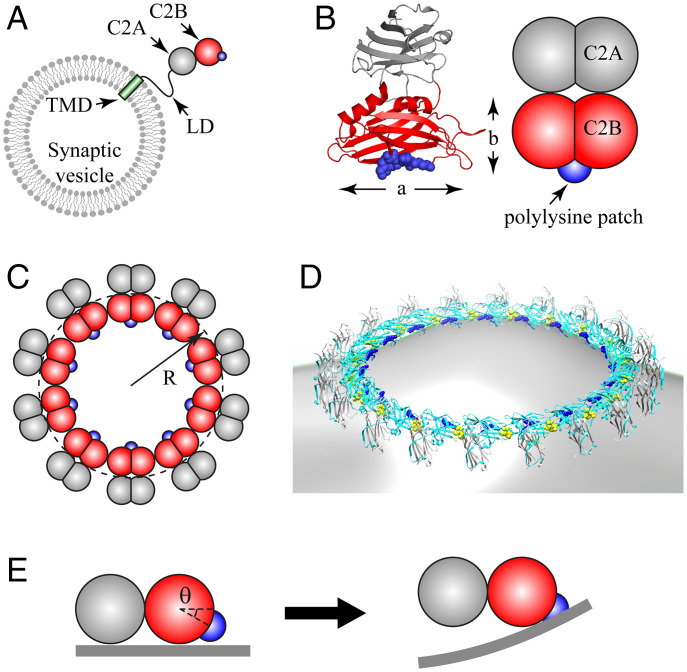
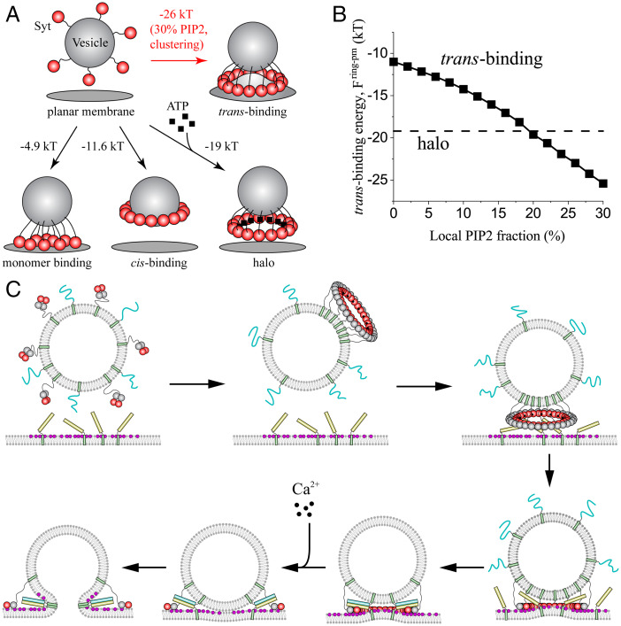
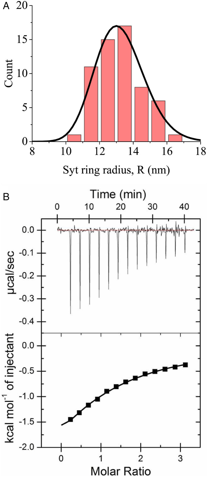
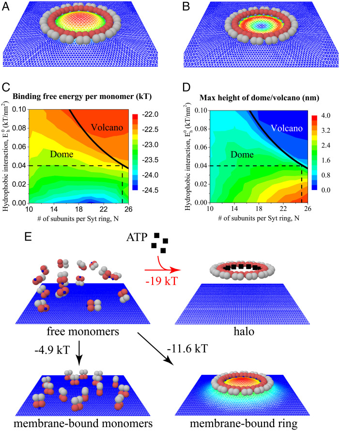
Synchronous release at neuronal synapses is accomplished by a machinery that senses calcium influx and fuses the synaptic vesicle and plasma membranes to release neurotransmitters. Previous studies suggested the calcium sensor synaptotagmin (Syt) is a facilitator of vesicle docking and both a facilitator and inhibitor of fusion. On phospholipid monolayers, the Syt C2AB domain spontaneously oligomerized into rings that are disassembled by Ca, suggesting Syt rings may clamp fusion as membrane-separating "washers" until Ca-mediated disassembly triggers fusion and release [J. Wang et al., 111, 13966-13971 (2014)].). Here, we combined mathematical modeling with experiment to measure the mechanical properties of Syt rings and to test this mechanism. Consistent with experimental results, the model quantitatively recapitulates observed Syt ring-induced dome and volcano shapes on phospholipid monolayers and predicts rings are stabilized by anionic phospholipid bilayers or bulk solution with ATP. The selected ring conformation is highly sensitive to membrane composition and bulk ATP levels, a property that may regulate vesicle docking and fusion in ATP-rich synaptic terminals. We find the Syt molecules hosted by a synaptic vesicle oligomerize into a halo, unbound from the vesicle, but in proximity to sufficiently phosphatidylinositol 4,5-bisphosphate (PIP2)-rich plasma membrane (PM) domains, the PM-bound trans Syt ring conformation is preferred. Thus, the Syt halo serves as landing gear for spatially directed docking at PIP2-rich sites that define the active zones of exocytotic release, positioning the Syt ring to clamp fusion and await calcium. Our results suggest the Syt ring is both a Ca-sensitive fusion clamp and a high-fidelity sensor for directed docking.
神经元突触的同步释放是通过一种机制实现的,该机制能够感知钙流入并融合突触囊泡和质膜以释放神经递质。先前的研究表明,钙传感器突触融合蛋白(Syt)是囊泡停靠的促进剂,同时也是融合的促进剂和抑制剂。在磷脂单层膜上,Syt C2AB 结构域自发寡聚形成环,这些环可被 Ca 解聚,这表明 Syt 环可能作为膜分离的“垫圈”夹住融合,直到 Ca 介导的解聚触发融合和释放[J. Wang 等人,111,13966-13971(2014)]。在这里,我们将数学建模与实验相结合,测量 Syt 环的力学特性并验证该机制。与实验结果一致,该模型定量再现了观察到的 Syt 环在磷脂单层膜上诱导的穹顶和火山形状,并预测了环被带负电荷的磷脂双层膜或含有 ATP 的体相溶液稳定。所选的环构象对膜组成和体相 ATP 水平高度敏感,这一特性可能调节富含 ATP 的突触末端的囊泡 docking 和融合。我们发现,突触囊泡中的 Syt 分子寡聚形成一个晕轮,与囊泡不结合,但靠近足够的磷脂酰肌醇 4,5-二磷酸(PIP2)丰富的质膜(PM)域,PM 结合的跨 Syt 环构象是首选的。因此,Syt 晕轮充当着着陆架,用于在富含 PIP2 的位点进行空间定向 docking,这些位点定义了胞吐释放的活性区,使 Syt 环能够夹住融合并等待 Ca。我们的结果表明,Syt 环既是一个 Ca 敏感的融合夹,也是一个用于定向 docking 的高保真传感器。