European Molecular Biology Laboratory (EMBL) Barcelona, Dr. Aiguader 88, 08003, Barcelona, Spain.
RIKEN Center for Biosystems Dynamics Research (RIKEN BDR), 2-2-3 Minatojima-minamimachi, Chuo-ku, 650-0047, Kobe, Japan.
Nat Commun. 2022 Sep 14;13(1):5400. doi: 10.1038/s41467-022-33115-0.
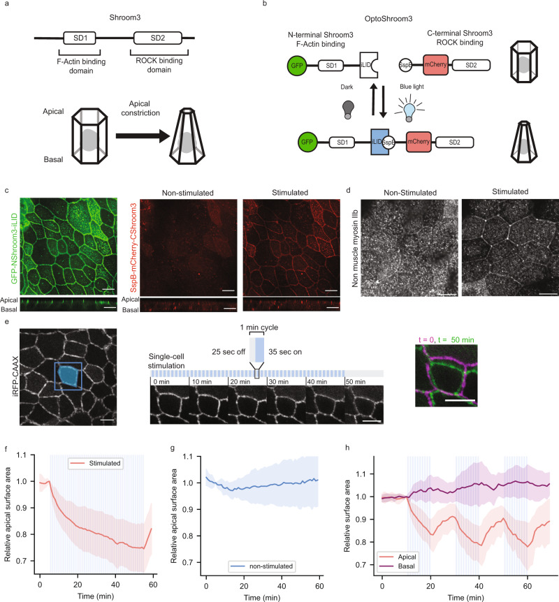
The emerging field of synthetic developmental biology proposes bottom-up approaches to examine the contribution of each cellular process to complex morphogenesis. However, the shortage of tools to manipulate three-dimensional (3D) shapes of mammalian tissues hinders the progress of the field. Here we report the development of OptoShroom3, an optogenetic tool that achieves fast spatiotemporal control of apical constriction in mammalian epithelia. Activation of OptoShroom3 through illumination in an epithelial Madin-Darby Canine Kidney (MDCK) cell sheet reduces the apical surface of the stimulated cells and causes displacements in the adjacent regions. Light-induced apical constriction provokes the folding of epithelial cell colonies on soft gels. Its application to murine and human neural organoids leads to thickening of neuroepithelia, apical lumen reduction in optic vesicles, and flattening in neuroectodermal tissues. These results show that spatiotemporal control of apical constriction can trigger several types of 3D deformation depending on the initial tissue context.
新兴的合成发育生物学领域提出了自下而上的方法来研究每个细胞过程对复杂形态发生的贡献。然而,用于操纵哺乳动物组织的三维(3D)形状的工具的缺乏阻碍了该领域的进展。在这里,我们报告了 OptoShroom3 的开发,这是一种光遗传学工具,可实现对哺乳动物上皮细胞的顶端收缩的快速时空控制。通过在上皮细胞 Madin-Darby 犬肾(MDCK)细胞片上的照明激活 OptoShroom3,可减少受刺激细胞的顶端表面,并导致相邻区域的位移。光诱导的顶端收缩会引起上皮细胞集落在软凝胶上的折叠。其在鼠和人神经类器官中的应用导致神经上皮的增厚、视泡中顶腔的减少以及神经外胚层组织的扁平化。这些结果表明,顶端收缩的时空控制可以根据初始组织背景触发几种类型的 3D 变形。