Immunology and Molecular Oncology Diagnostics, Veneto Institute of Oncology IOV-IRCCS, Via Gattamelata 64, 35128, Padova, Italy.
Department of Surgery, Oncology and Gastroenterology, University of Padova, Padova, Italy.
J Exp Clin Cancer Res. 2022 Sep 20;41(1):279. doi: 10.1186/s13046-022-02481-4.
Immune checkpoint inhibitors (ICI) are approved for treatment of recurrent or metastatic oropharyngeal head and neck squamous cell carcinoma in the first- and second-line settings. However, only 15-20% of patients benefit from this treatment, a feature increasingly ascribed to the peculiar characteristics of the tumor immune microenvironment (TIME).
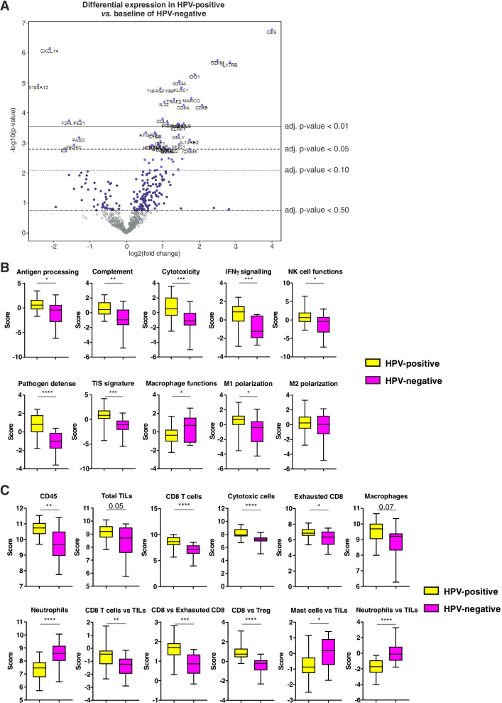
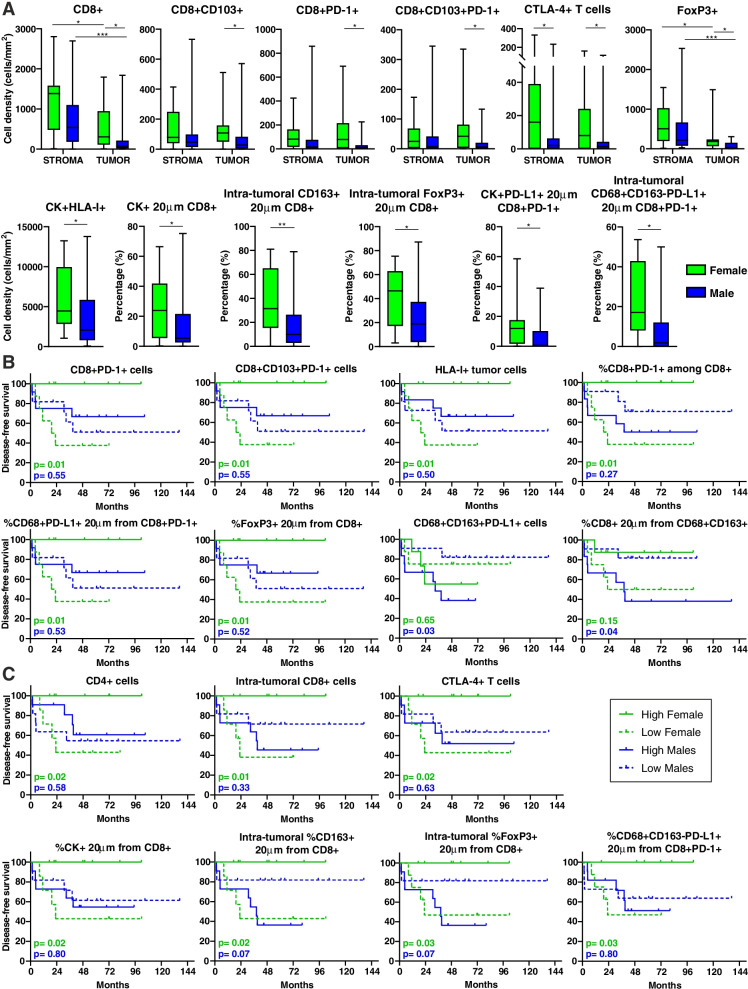
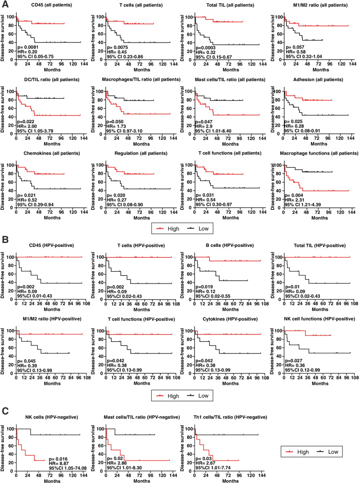
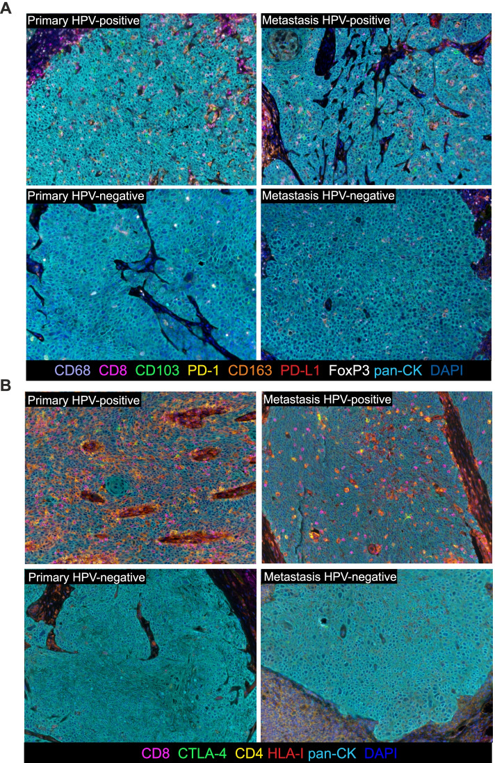
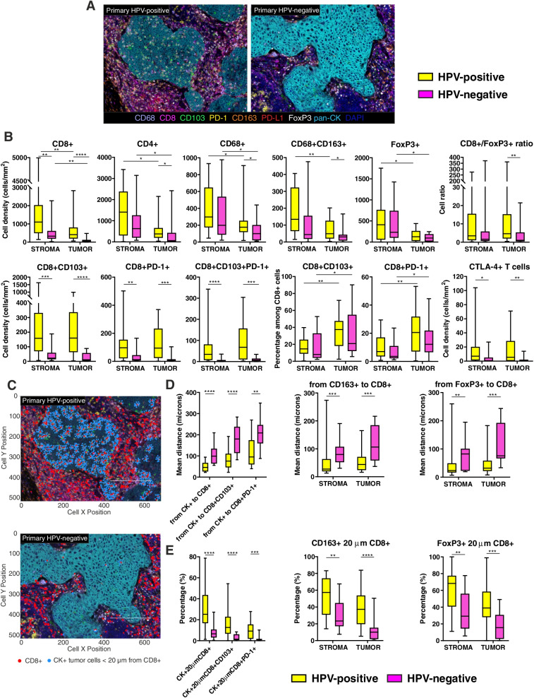
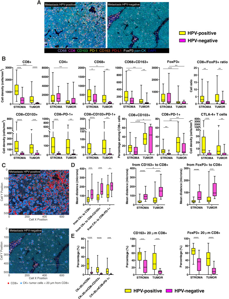
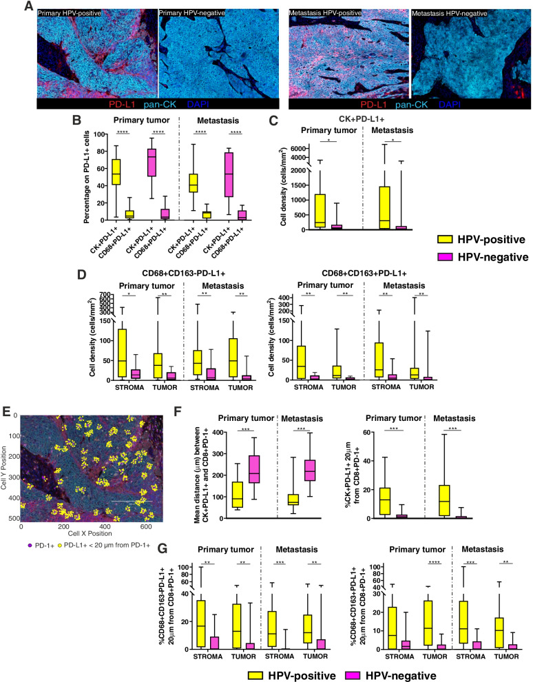
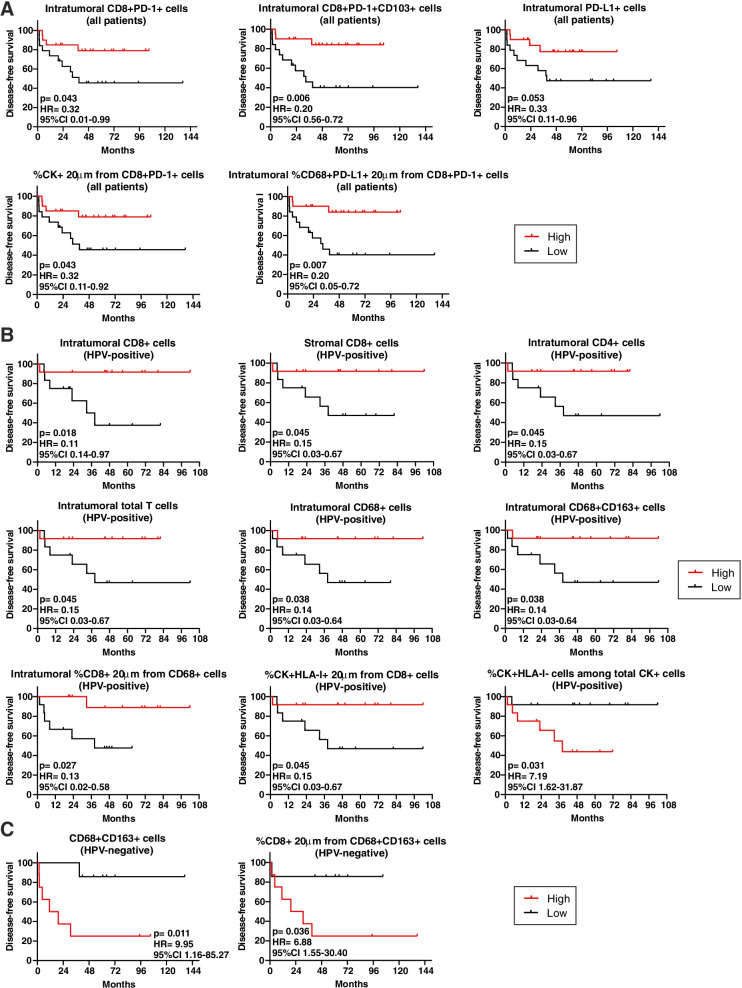
Immune-related gene expression profiling (GEP) and multiplex immunofluorescence (mIF) including spatial proximity analysis, were used to characterize the TIME of 39 treatment-naïve oropharyngeal squamous cell carcinomas (OPSCC) and the corresponding lymph node metastases. GEP and mIF results were correlated with disease-free survival (DFS). HPV-positive tumors disclosed a stronger activation of several immune signalling pathways, as well as a higher expression of genes related to total tumor-infiltrating lymphocytes, CD8 T cells, cytotoxic cells and exhausted CD8 cells, than HPV-negative patients. Accordingly, mIF revealed that HPV-positive lesions were heavily infiltrated as compared to HPV-negative counterparts, with a higher density of T cells and checkpoint molecules. CD8+ T cells appeared in closer proximity to tumor cells, CD163+ macrophages and FoxP3+ cells in HPV-positive primary tumors, and related metastases. In HPV-positive lesions, PD-L1 expression was increased as compared to HPV-negative samples, and PD-L1+ tumor cells and macrophages were closer to PD-1+ cytotoxic T lymphocytes. Considering the whole cohort, a positive correlation was observed between DFS and higher levels of activating immune signatures and T cell responses, higher density of PD-1+ T cells and their closer proximity to tumor cells or PD-L1+ macrophages. HPV-positive patients with higher infiltration of T cells and macrophages had a longer DFS, while CD163+ macrophages had a negative role in prognosis of HPV-negative patients.
Our results suggest that checkpoint expression may reflect an ongoing antitumor immune response. Thus, these observations provide the rationale for the incorporation of ICI in the loco-regional therapy strategies for patients with heavily infiltrated treatment-naïve OPSCC, and for the combination of ICI with tumor-specific T cell response inducers or TAM modulators for the "cold" OPSCC counterparts.
免疫检查点抑制剂(ICI)已被批准用于治疗一线和二线复发性或转移性或口咽头颈部鳞状细胞癌。然而,只有 15-20%的患者从中受益,这一特征越来越归因于肿瘤免疫微环境(TIME)的特殊特征。
采用免疫相关基因表达谱(GEP)和多重免疫荧光(mIF),包括空间邻近分析,对 39 例未经治疗的口咽鳞状细胞癌(OPSCC)及其相应的淋巴结转移进行 TIME 特征分析。GEP 和 mIF 结果与无病生存期(DFS)相关。HPV 阳性肿瘤显示出几种免疫信号通路的更强激活,以及与总肿瘤浸润淋巴细胞、CD8 T 细胞、细胞毒性细胞和耗竭 CD8 细胞相关的基因表达更高,而 HPV 阴性患者则较低。因此,mIF 显示 HPV 阳性病变比 HPV 阴性病变浸润更严重,T 细胞和检查点分子密度更高。CD8+T 细胞与肿瘤细胞、CD163+巨噬细胞和 FoxP3+细胞在 HPV 阳性原发性肿瘤及其相关转移中更接近。在 HPV 阳性病变中,与 HPV 阴性样本相比,PD-L1 表达增加,PD-L1+肿瘤细胞和巨噬细胞更接近 PD-1+细胞毒性 T 淋巴细胞。考虑到整个队列,DFS 与更高水平的激活免疫特征和 T 细胞反应、更高密度的 PD-1+T 细胞及其与肿瘤细胞或 PD-L1+巨噬细胞的更接近呈正相关。T 细胞和巨噬细胞浸润较高的 HPV 阳性患者 DFS 较长,而 CD163+巨噬细胞在 HPV 阴性患者的预后中起负面作用。
我们的研究结果表明,检查点表达可能反映了持续的抗肿瘤免疫反应。因此,这些观察结果为将 ICI 纳入局部区域治疗策略提供了依据,用于治疗浸润性强的初治 OPSCC 患者,以及为“冷”OPSCC 患者联合 ICI 和肿瘤特异性 T 细胞反应诱导剂或 TAM 调节剂。