• 文献检索
  • 文档翻译
  • 深度研究
  • 学术资讯
  • Suppr Zotero 插件Zotero 插件
  • 邀请有礼
  • 套餐&价格
  • 历史记录
应用&插件
Suppr Zotero 插件Zotero 插件浏览器插件Mac 客户端Windows 客户端微信小程序
定价
高级版会员购买积分包购买API积分包
服务
文献检索文档翻译深度研究API 文档MCP 服务
关于我们
关于 Suppr公司介绍联系我们用户协议隐私条款
关注我们

Suppr 超能文献

核心技术专利:CN118964589B侵权必究
粤ICP备2023148730 号-1Suppr @ 2026

文献检索

告别复杂PubMed语法,用中文像聊天一样搜索,搜遍4000万医学文献。AI智能推荐,让科研检索更轻松。

立即免费搜索

文件翻译

保留排版,准确专业,支持PDF/Word/PPT等文件格式,支持 12+语言互译。

免费翻译文档

深度研究

AI帮你快速写综述,25分钟生成高质量综述,智能提取关键信息,辅助科研写作。

立即免费体验

使用光学相干断层扫描技术对比圆锥角膜与正常眼的角膜总厚度分布模式

Distribution Pattern of Total Corneal Thickness in Keratoconus versus Normal Eyes Using an Optical Coherence Tomography.

作者信息

Hashemi Hassan, Heidari Zahra, Mohammadpour Mehrdad, Momeni-Moghaddam Hamed, Khabazkhoob Mehdi

机构信息

Noor Ophthalmology Research Center, Noor Eye Hospital, Tehran, Iran.

Department of Ophthalmology, Bu-Ali Sina Hospital, Mazandaran University of Medical Sciences, Sari, Iran.

出版信息

J Curr Ophthalmol. 2022 Jul 26;34(2):216-222. doi: 10.4103/joco.joco_198_21. eCollection 2022 Apr-Jun.

DOI:10.4103/joco.joco_198_21
PMID:36147258
原文链接:https://pmc.ncbi.nlm.nih.gov/articles/PMC9487006/
Abstract

PURPOSE

To evaluate the total corneal thickness distribution pattern using a high-resolution spectral-domain optical coherence tomography (HR SD-OCT) for distinguishing normal eyes from keratoconus (KCN).

METHODS

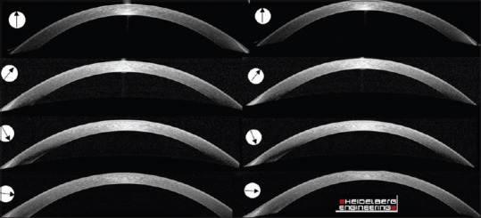
One hundred and forty-four patients were enrolled in three groups (55 normal, 45 mild KCN, and 44 moderate-to-severe KCN eyes) in this prospective diagnostic test study. Total corneal thickness was measured in 8 semi-meridians using HR SD-OCT (Heidelberg Engineering, Heidelberg, Germany) in 5 and 7 mm zones. The central corneal thickness (CCT), corneal focal thinning (minimum thickness [Min], min minus median and maximum [Min-Med, Min-Max]), and asymmetry indices (inferior minus superior [I-S] and supranasal minus infratemporal [SN-IT]) were calculated. One-way analysis of variance and the area under the receiver operating characteristic curve (AUC) were used for the analysis.

RESULTS

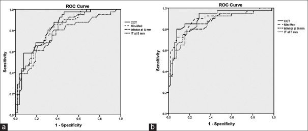
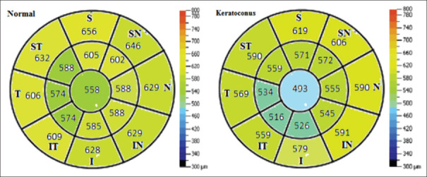
Thinner CCT, lower Min thickness, more negative Min-Max, Min-Med, and greater I-S and SN-IT were found in KCN eyes compared to the control group ( < 0.001). The inferior and IT semi-meridians were the thinnest locations in KCN cases in the 5 mm central zone ( < 0.001). CCT followed by Min-Med had the highest discriminative ability for differentiating mild KCN (AUC, sensitivity and specificity: 0.822, 87.0%, 60.37% and 0.805, 82.93%, 66.0%, respectively) and moderate-to-severe KCN (0.902, 87.82%, 73.08% and 0.892, 85.37%, and 78.85%, respectively) from normal corneas.

CONCLUSION

The inferior and IT sectors of the cornea with the largest thickness changes in the 5 mm zone are the most common thinning sites in keratoconic corneas, and CCT and Min-Med are the most sensitive indices for the diagnosis of KCN.

摘要

目的

使用高分辨率光谱域光学相干断层扫描(HR SD-OCT)评估全角膜厚度分布模式,以区分正常眼与圆锥角膜(KCN)。

方法

在这项前瞻性诊断试验研究中,144例患者被纳入三组(55只正常眼、45只轻度KCN眼和44只中重度KCN眼)。使用HR SD-OCT(德国海德堡海德堡工程公司)在5毫米和7毫米区域的8个半子午线上测量全角膜厚度。计算中央角膜厚度(CCT)、角膜局部变薄(最小厚度[Min]、最小厚度减去中位数和最大值[Min-Med,Min-Max])以及不对称指数(下方减去上方[I-S]和鼻上侧减去颞下侧[SN-IT])。采用单因素方差分析和受试者操作特征曲线下面积(AUC)进行分析。

结果

与对照组相比,KCN眼中CCT更薄、Min厚度更低、Min-Max和Min-Med更负,且I-S和SN-IT更大(<0.001)。在5毫米中央区域,下方和颞下侧半子午线是KCN病例中最薄的部位(<0.001)。对于区分轻度KCN(AUC、敏感性和特异性分别为:0.822、87.0%、60.37%)和中重度KCN(0.902、87.82%、73.08%)与正常角膜,CCT其次是Min-Med具有最高的鉴别能力(分别为0.805、82.93%、66.0%和0.892、85.37%、78.85%)。

结论

在5毫米区域厚度变化最大的角膜下方和颞下侧区域是圆锥角膜最常见的变薄部位,CCT和Min-Med是诊断KCN最敏感的指标。

https://cdn.ncbi.nlm.nih.gov/pmc/blobs/36cb/9487006/31e7115c78c4/JCO-34-216-g003.jpg
https://cdn.ncbi.nlm.nih.gov/pmc/blobs/36cb/9487006/47fb43d50af4/JCO-34-216-g001.jpg
https://cdn.ncbi.nlm.nih.gov/pmc/blobs/36cb/9487006/a2a1959acdcd/JCO-34-216-g002.jpg
https://cdn.ncbi.nlm.nih.gov/pmc/blobs/36cb/9487006/31e7115c78c4/JCO-34-216-g003.jpg
https://cdn.ncbi.nlm.nih.gov/pmc/blobs/36cb/9487006/47fb43d50af4/JCO-34-216-g001.jpg
https://cdn.ncbi.nlm.nih.gov/pmc/blobs/36cb/9487006/a2a1959acdcd/JCO-34-216-g002.jpg
https://cdn.ncbi.nlm.nih.gov/pmc/blobs/36cb/9487006/31e7115c78c4/JCO-34-216-g003.jpg

相似文献

1
Distribution Pattern of Total Corneal Thickness in Keratoconus versus Normal Eyes Using an Optical Coherence Tomography.使用光学相干断层扫描技术对比圆锥角膜与正常眼的角膜总厚度分布模式
J Curr Ophthalmol. 2022 Jul 26;34(2):216-222. doi: 10.4103/joco.joco_198_21. eCollection 2022 Apr-Jun.
2
[The study of corneal epithelial change in keratoconic eyes with Fourier-domain optical coherence tomographic pachymetry].[傅里叶域光学相干断层扫描测厚法对圆锥角膜眼角膜上皮变化的研究]
Zhonghua Yan Ke Za Zhi. 2014 Sep;50(9):665-70.
3
Corneal layer thickness in keratoconus using optical coherence tomography.角膜层厚度在圆锥角膜的光学相干断层扫描。
Clin Exp Optom. 2024 Jan;107(1):32-39. doi: 10.1080/08164622.2023.2201369. Epub 2023 Apr 30.
4
OCT-derived comparison of corneal thickness distribution and asymmetry differences between normal and keratoconic eyes.基于光学相干断层扫描(OCT)的正常眼与圆锥角膜眼之间角膜厚度分布及不对称性差异的比较
Cornea. 2014 Dec;33(12):1274-81. doi: 10.1097/ICO.0000000000000275.
5
Corneal epithelial thickness mapping by Fourier-domain optical coherence tomography in normal and keratoconic eyes.傅里叶域光学相干断层扫描对正常眼和圆锥角膜眼的角膜上皮厚度测绘。
Ophthalmology. 2012 Dec;119(12):2425-33. doi: 10.1016/j.ophtha.2012.06.023. Epub 2012 Aug 20.
6
Keratoconus diagnosis with optical coherence tomography pachymetry mapping.基于光学相干断层扫描测厚成像的圆锥角膜诊断
Ophthalmology. 2008 Dec;115(12):2159-66. doi: 10.1016/j.ophtha.2008.08.004. Epub 2008 Nov 5.
7
Corneal epithelial thickness mapping using Fourier-domain optical coherence tomography for detection of form fruste keratoconus.使用傅里叶域光学相干断层扫描进行角膜上皮厚度测绘以检测forme fruste圆锥角膜。
J Cataract Refract Surg. 2015 Apr;41(4):812-20. doi: 10.1016/j.jcrs.2014.06.043.
8
Distinguishing Highly Asymmetric Keratoconus Eyes Using Combined Scheimpflug and Spectral-Domain OCT Analysis.使用联合 Scheimpflug 和频域 OCT 分析区分高度非对称圆锥角膜眼。
Ophthalmology. 2018 Dec;125(12):1862-1871. doi: 10.1016/j.ophtha.2018.06.020. Epub 2018 Jul 25.
9
Optical coherence tomography-derived corneal thickness asymmetry indices: clinical reference study of normal eyes.光学相干断层扫描衍生的角膜厚度不对称指数:正常眼的临床参考研究
J Cataract Refract Surg. 2014 Oct;40(10):1603-9. doi: 10.1016/j.jcrs.2014.01.041. Epub 2014 Aug 28.
10
Subclinical keratoconus detection by pattern analysis of corneal and epithelial thickness maps with optical coherence tomography.通过光学相干断层扫描对角膜和上皮厚度图进行模式分析检测亚临床圆锥角膜
J Cataract Refract Surg. 2016 Feb;42(2):284-95. doi: 10.1016/j.jcrs.2015.09.021.

引用本文的文献

1
Digital transformation of care for keratoconus patients: ML modeling structural outcomes of corneal collagen cross-linking.圆锥角膜患者护理的数字化转型:角膜胶原交联的机器学习建模结构结果
Front Med (Lausanne). 2025 Jun 4;12:1462653. doi: 10.3389/fmed.2025.1462653. eCollection 2025.
2
Topographic factors associated with anterior chamber angle narrowing in patients with keratoconus.圆锥角膜患者前房角狭窄相关的地形学因素。
Int J Ophthalmol. 2025 Jan 18;18(1):67-73. doi: 10.18240/ijo.2025.01.07. eCollection 2025.
3
Detecting Keratoconus in Adolescents with Anterior Segment Optical Coherence Tomography.

本文引用的文献

1
A Case-Control Study of Keratoconus Risk Factors.圆锥角膜危险因素的病例对照研究。
Cornea. 2020 Jun;39(6):697-701. doi: 10.1097/ICO.0000000000002283.
2
Optical coherence tomography imaging in keratoconus.圆锥角膜的光学相干断层扫描成像
Clin Exp Optom. 2019 May;102(3):218-223. doi: 10.1111/cxo.12874. Epub 2019 Feb 22.
3
Safety and Efficacy of Sequential Intracorneal Ring Segment Implantation and Cross-linking in Pediatric Keratoconus.小儿圆锥角膜序贯式角膜内环植入术与交联术的安全性和有效性
使用眼前节光学相干断层扫描技术检测青少年圆锥角膜
J Ophthalmol. 2024 Jun 7;2024:6655217. doi: 10.1155/2024/6655217. eCollection 2024.
4
Analysis of Corneal Biomechanical Properties in Different Keratotopographic Patterns of Keratoconus.圆锥角膜不同角膜地形图模式下的角膜生物力学特性分析
J Curr Ophthalmol. 2024 Mar 29;35(3):249-258. doi: 10.4103/joco.joco_83_23. eCollection 2023 Jul-Sep.
5
Diagnostic Accuracy of Corneal and Epithelial Thickness Map Parameters to Detect Keratoconus and Suspect Keratoconus.用于检测圆锥角膜和疑似圆锥角膜的角膜及上皮厚度图参数的诊断准确性
J Ophthalmol. 2023 Oct 6;2023:6677932. doi: 10.1155/2023/6677932. eCollection 2023.
Am J Ophthalmol. 2017 Jun;178:51-57. doi: 10.1016/j.ajo.2017.03.016. Epub 2017 Mar 22.
4
Comparison of In Vivo Confocal Microscopy, Ultrasonic Pachymetry, and Scheimpflug Topography for Measuring Central Corneal Thickness.体内共焦显微镜、超声测厚法和眼前节分析系统测量中央角膜厚度的比较
JAMA Ophthalmol. 2016 Sep 1;134(9):1057-9. doi: 10.1001/jamaophthalmol.2016.2183.
5
Assessment of Corneal Epithelial Thickness in Asymmetric Keratoconic Eyes and Normal Eyes Using Fourier Domain Optical Coherence Tomography.使用傅里叶域光学相干断层扫描技术评估非对称性圆锥角膜眼和正常眼的角膜上皮厚度
J Ophthalmol. 2016;2016:5697343. doi: 10.1155/2016/5697343. Epub 2016 Jun 9.
6
Evaluation of Total Corneal Thickness and Corneal Layers With Spectral-Domain Optical Coherence Tomography.利用频域光学相干断层扫描技术评估角膜总厚度及各层结构
J Refract Surg. 2016 Jan;32(1):27-32. doi: 10.3928/1081597X-20151207-03.
7
Global Survey of Corneal Transplantation and Eye Banking.全球角膜移植与眼库调查
JAMA Ophthalmol. 2016 Feb;134(2):167-73. doi: 10.1001/jamaophthalmol.2015.4776.
8
Comparison of Fourier-Domain and Time-Domain Optical Coherence Tomography in the Measurement of Thinnest Corneal Thickness in Keratoconus.傅里叶域与时域光学相干断层扫描在圆锥角膜最薄角膜厚度测量中的比较
J Ophthalmol. 2015;2015:402925. doi: 10.1155/2015/402925. Epub 2015 Jun 25.
9
Corneal epithelial thickness mapping using Fourier-domain optical coherence tomography for detection of form fruste keratoconus.使用傅里叶域光学相干断层扫描进行角膜上皮厚度测绘以检测forme fruste圆锥角膜。
J Cataract Refract Surg. 2015 Apr;41(4):812-20. doi: 10.1016/j.jcrs.2014.06.043.
10
Comparison of central corneal thickness measurements using optical low-coherence reflectometry, Fourier domain optical coherence tomography, and Scheimpflug camera.使用光学低相干反射测量法、傅里叶域光学相干断层扫描术和Scheimpflug相机测量中央角膜厚度的比较。
Arq Bras Oftalmol. 2014 Nov-Dec;77(6):345-50. doi: 10.5935/0004-2749.20140087.