• 文献检索
  • 文档翻译
  • 深度研究
  • 学术资讯
  • Suppr Zotero 插件Zotero 插件
  • 邀请有礼
  • 套餐&价格
  • 历史记录
应用&插件
Suppr Zotero 插件Zotero 插件浏览器插件Mac 客户端Windows 客户端微信小程序
定价
高级版会员购买积分包购买API积分包
服务
文献检索文档翻译深度研究API 文档MCP 服务
关于我们
关于 Suppr公司介绍联系我们用户协议隐私条款
关注我们

Suppr 超能文献

核心技术专利:CN118964589B侵权必究
粤ICP备2023148730 号-1Suppr @ 2026

文献检索

告别复杂PubMed语法,用中文像聊天一样搜索,搜遍4000万医学文献。AI智能推荐,让科研检索更轻松。

立即免费搜索

文件翻译

保留排版,准确专业,支持PDF/Word/PPT等文件格式,支持 12+语言互译。

免费翻译文档

深度研究

AI帮你快速写综述,25分钟生成高质量综述,智能提取关键信息,辅助科研写作。

立即免费体验

5T与3T的肾脏成像:一项比较研究。

Renal imaging at 5 T versus 3 T: a comparison study.

作者信息

Zheng Liyun, Yang Chun, Sheng Ruofan, Dai Yongming, Zeng Mengsu

机构信息

Shanghai Institute of Medical Imaging, Shanghai, China.

Department of Radiology, Zhongshan Hospital, Fudan University, No. 180 Fenglin Road, Xuhui District, Shanghai, 20032, China.

出版信息

Insights Imaging. 2022 Sep 24;13(1):155. doi: 10.1186/s13244-022-01290-9.

DOI:10.1186/s13244-022-01290-9
PMID:36153471
原文链接:https://pmc.ncbi.nlm.nih.gov/articles/PMC9509503/
Abstract

BACKGROUND

Recently, a whole-body 5 T MRI scanner was developed to open the door of abdominal imaging at high-field strength. This prospective study aimed to evaluate the feasibility of renal imaging at 5 T and compare the image quality, potential artifacts, and contrast ratios with 3 T.

METHODS

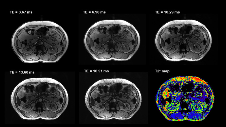
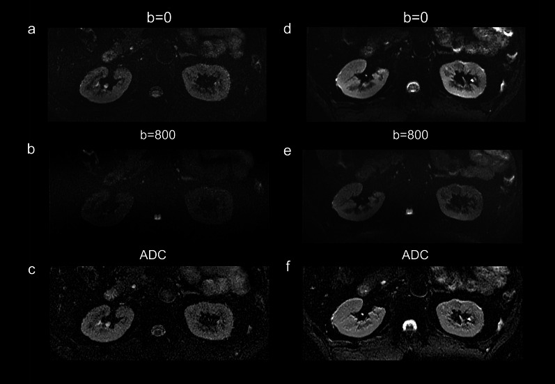
Forty healthy volunteers underwent MRI examination both at 3 T and 5 T. MRI sequences included T1-weighted gradient-echo (GRE), T2-weighted fast spin echo, diffusion-weighted imaging, and multi-echo GRE T2* mapping. Image quality and presence of artifacts were assessed for all sequences using four-point scales. For anatomical imaging, the signal-to-noise ratio (SNR) and contrast ratio (CR) of abdomen organ tissues were calculated. Besides, for functional imaging, the contrast-to-noise ratio of cortex/medulla was calculated. Wilcoxon signed rank-sum test was used to compare the visual evaluation scores and quantitative measurements between 3 and 5 T images.

RESULTS

Compared to 3 T examination, T1-weighted sequence at 5 T showed significantly better image quality with higher conspicuity of the renal veins and arteries, and comparable artifacts. Image quality was comparable between both field strengths on T2-weighted images, whereas a significantly higher level of artifacts was observed at 5 T. Besides, 5 T MRI contributed to higher SNR and CR for abdomen organ tissues. For functional imaging, 5 T MRI showed improved corticomedullar discrimination. There was no significant difference between apparent diffusion coefficient of renal at 3 T and 5 T, while 5 T MRI resulted in significantly shorter T2* values in both cortex and medulla.

CONCLUSIONS

5 T MRI provides anatomical and functional images of the kidney with sufficient image quality.

摘要

背景

最近,一台全身5T磁共振成像(MRI)扫描仪被开发出来,开启了高场强腹部成像的大门。这项前瞻性研究旨在评估5T下肾脏成像的可行性,并将图像质量、潜在伪影以及对比率与3T进行比较。

方法

40名健康志愿者分别接受了3T和5T的MRI检查。MRI序列包括T1加权梯度回波(GRE)、T2加权快速自旋回波、扩散加权成像以及多回波GRE T2*映射。使用四点量表对所有序列的图像质量和伪影情况进行评估。对于解剖成像,计算腹部器官组织的信噪比(SNR)和对比率(CR)。此外,对于功能成像,计算皮质/髓质的对比噪声比。采用Wilcoxon符号秩和检验来比较3T和5T图像之间的视觉评估分数和定量测量结果。

结果

与3T检查相比,5T下的T1加权序列显示出明显更好的图像质量,肾动静脉显示更清晰,伪影情况相当。T2加权图像上两种场强的图像质量相当,但5T时观察到的伪影水平明显更高。此外,5T MRI使腹部器官组织的SNR和CR更高。对于功能成像,5T MRI显示皮质髓质分辨能力有所提高。3T和5T时肾脏的表观扩散系数之间无显著差异,但5T MRI使皮质和髓质的T2*值均显著缩短。

结论

5T MRI能够提供具有足够图像质量的肾脏解剖和功能图像。

https://cdn.ncbi.nlm.nih.gov/pmc/blobs/5126/9509503/f49b9bd82525/13244_2022_1290_Fig4_HTML.jpg
https://cdn.ncbi.nlm.nih.gov/pmc/blobs/5126/9509503/fff73ecde01b/13244_2022_1290_Fig1_HTML.jpg
https://cdn.ncbi.nlm.nih.gov/pmc/blobs/5126/9509503/19847b719adf/13244_2022_1290_Fig2_HTML.jpg
https://cdn.ncbi.nlm.nih.gov/pmc/blobs/5126/9509503/68a124b6200b/13244_2022_1290_Fig3_HTML.jpg
https://cdn.ncbi.nlm.nih.gov/pmc/blobs/5126/9509503/f49b9bd82525/13244_2022_1290_Fig4_HTML.jpg

相似文献

1
Renal imaging at 5 T versus 3 T: a comparison study.5T与3T的肾脏成像:一项比较研究。
Insights Imaging. 2022 Sep 24;13(1):155. doi: 10.1186/s13244-022-01290-9.
2
Comparison of multi-echo and single-echo gradient-recalled echo sequences for SPIO-enhanced liver MRI at 3 T.多回波与单次激发梯度回波序列在 3.0T 场强下用于 SPIO 增强肝脏 MRI 的对比研究。
Clin Radiol. 2010 Nov;65(11):916-23. doi: 10.1016/j.crad.2010.07.003.
3
Comparison of HASTE with multiple signal averaging versus conventional turbo spin echo sequence: a new option for T2-weighted MRI of the female pelvis.HASTE 与多信号平均法与常规 turbo 自旋回波序列的比较:女性盆腔 T2 加权 MRI 的新选择。
Eur Radiol. 2020 Jun;30(6):3245-3253. doi: 10.1007/s00330-020-06686-z. Epub 2020 Feb 16.
4
Hepatocellular lesions with increased iron uptake on superparamagnetic iron oxide-enhanced magnetic resonance imaging in cirrhosis or chronic hepatitis: comparison of four magnetic resonance sequences for lesion conspicuity.肝硬化或慢性肝炎患者中,超顺磁性氧化铁增强磁共振成像显示铁摄取增加的肝细胞病变:四种磁共振序列对病变显示清晰度的比较
Magn Reson Imaging. 2009 Jul;27(6):801-6. doi: 10.1016/j.mri.2008.11.006. Epub 2009 Jan 13.
5
Fast multiplanar spoiled gradient-recalled imaging of the liver: pulse sequence optimization and comparison with spin-echo MR imaging.肝脏快速多平面扰相梯度回波成像:脉冲序列优化及与自旋回波磁共振成像的比较
AJR Am J Roentgenol. 1993 Mar;160(3):501-9. doi: 10.2214/ajr.160.3.8381572.
6
Comparison of T1-weighted in- and out-of-phase single shot magnetization-prepared gradient-recalled-echo with three-dimensional gradient-recalled-echo at 3.0 Tesla: preliminary observations in abdominal studies.3.0T 磁共振 T1WI 同反相位单次激发磁化准备梯度回波与三维梯度回波序列在腹部的初步应用比较
J Magn Reson Imaging. 2012 May;35(5):1187-95. doi: 10.1002/jmri.23518. Epub 2011 Nov 29.
7
Contrast-Enhanced T1-Weighted Head and Neck MRI: Prospective Intraindividual Image Quality Comparison of Spiral GRE, Cartesian GRE, and Cartesian TSE Sequences.对比增强T1加权头颈部磁共振成像:螺旋梯度回波、笛卡尔梯度回波和笛卡尔快速自旋回波序列的前瞻性个体内图像质量比较
AJR Am J Roentgenol. 2022 Jan;218(1):132-139. doi: 10.2214/AJR.21.26413. Epub 2021 Aug 18.
8
Dynamic contrast-enhanced renal MRI at 7 Tesla: preliminary results.7 特斯拉下动态对比增强肾 MRI:初步结果。
Invest Radiol. 2011 Jul;46(7):425-33. doi: 10.1097/RLI.0b013e31820e1467.
9
Simultaneous Multislice Diffusion-Weighted Imaging of the Kidney: A Systematic Analysis of Image Quality.肾脏的同时多层扩散加权成像:图像质量的系统分析
Invest Radiol. 2017 Mar;52(3):163-169. doi: 10.1097/RLI.0000000000000323.
10
Artifact-robust diffusion-weighted whole-body imaging with background suppression at 3 T using improved turbo spin-echo diffusion-weighted imaging.利用改进的快速自旋回波扩散加权成像在3T下进行具有背景抑制的伪影稳健型扩散加权全身成像。
Br J Radiol. 2019 Feb;92(1094):20180489. doi: 10.1259/bjr.20180489. Epub 2018 Nov 14.

引用本文的文献

1
Application of AI-assisted compressed sensing in high resolution Gd-EOB-DTPA-enhanced MRI during hepatobiliary phase at 5.0 T.人工智能辅助压缩感知在5.0T肝胆期高分辨率钆塞酸二钠增强磁共振成像中的应用
Quant Imaging Med Surg. 2025 Aug 1;15(8):6852-6867. doi: 10.21037/qims-2025-264. Epub 2025 Jul 25.
2
Diff5T: Benchmarking human brain diffusion MRI with an extensive 5.0 Tesla k-space and spatial dataset.Diff5T:使用广泛的5.0特斯拉k空间和空间数据集对人类脑扩散磁共振成像进行基准测试。
Sci Data. 2025 Aug 4;12(1):1352. doi: 10.1038/s41597-025-05640-2.
3
Performance Evaluation of a Custom-Designed Contrast Media Injector in a 5-Tesla MRI Environment.

本文引用的文献

1
Preliminary Experience of 5.0 T Higher Field Abdominal Diffusion-Weighted MRI: Agreement of Apparent Diffusion Coefficient With 3.0 T Imaging.5.0T 高场腹部扩散加权 MRI 的初步经验:表观扩散系数与 3.0T 成像的一致性。
J Magn Reson Imaging. 2022 Oct;56(4):1009-1017. doi: 10.1002/jmri.28097. Epub 2022 Feb 4.
2
Emerging Use of Ultra-High-Field 7T MRI in the Study of Intracranial Vascularity: State of the Field and Future Directions.超高场 7T MRI 在颅内血管研究中的新兴应用:现状与未来方向。
AJNR Am J Neuroradiol. 2020 Jan;41(1):2-9. doi: 10.3174/ajnr.A6344. Epub 2019 Dec 26.
3
Magnetic Resonance Imaging of the Musculoskeletal System at 7T: Morphological Imaging and Beyond.
在5特斯拉磁共振成像环境中对定制设计的造影剂注射器的性能评估
Bioengineering (Basel). 2025 May 25;12(6):566. doi: 10.3390/bioengineering12060566.
4
Diagnostic ultrasound characteristics of Morel-Lavallée lesions: Case and dataset analysis.莫雷尔-拉瓦利埃损伤的诊断超声特征:病例与数据集分析
Medicine (Baltimore). 2025 Jun 20;104(25):e42906. doi: 10.1097/MD.0000000000042906.
5
Magnetic resonance imaging of renal oxygenation.肾脏氧合的磁共振成像。
Nat Rev Nephrol. 2025 Apr 23. doi: 10.1038/s41581-025-00956-z.
6
Quo Vadis MRI?MRI 何去何从?
Z Med Phys. 2025 Feb;35(1):3-5. doi: 10.1016/j.zemedi.2024.12.002. Epub 2025 Jan 11.
7
Magnetic resonance cholangiopancreatography at 5.0 T: quantitative and qualitative comparison with 3.0 T.5.0 T磁共振胰胆管造影:与3.0 T的定量和定性比较
BMC Med Imaging. 2024 Dec 5;24(1):331. doi: 10.1186/s12880-024-01512-0.
8
Improved Detection of Target Metabolites in Brain Tumors with Intermediate TE, High SNR, and High Bandwidth Spin-Echo Sequence at 5T.5T 下 TE 适中、SNR 高、带宽高的自旋回波序列可提高脑肿瘤靶代谢物的检测。
AJNR Am J Neuroradiol. 2024 Apr 8;45(4):461-467. doi: 10.3174/ajnr.A8150.
7T 磁共振成像在肌肉骨骼系统中的应用:形态学成像及其他
Top Magn Reson Imaging. 2019 Jun;28(3):125-135. doi: 10.1097/RMR.0000000000000205.
4
Clinical 7 T MRI: Are we there yet? A review about magnetic resonance imaging at ultra-high field.临床7T磁共振成像:我们到那一步了吗?关于超高场磁共振成像的综述
Br J Radiol. 2019 Feb;92(1094):20180492. doi: 10.1259/bjr.20180492. Epub 2018 Nov 1.
5
Functional Evaluation of Transplanted Kidneys with Reduced Field-of-View Diffusion-Weighted Imaging at 3T.3T 下小视野扩散加权成像评估移植肾功能。
Korean J Radiol. 2018 Mar-Apr;19(2):201-208. doi: 10.3348/kjr.2018.19.2.201. Epub 2018 Feb 22.
6
1.5 versus 3 versus 7 Tesla in abdominal MRI: A comparative study.腹部磁共振成像中1.5特斯拉、3特斯拉与7特斯拉的比较研究
PLoS One. 2017 Nov 10;12(11):e0187528. doi: 10.1371/journal.pone.0187528. eCollection 2017.
7
7T: Physics, safety, and potential clinical applications.7T:物理特性、安全性和潜在临床应用。
J Magn Reson Imaging. 2017 Dec;46(6):1573-1589. doi: 10.1002/jmri.25723. Epub 2017 Apr 3.
8
7T MRI for neurodegenerative dementias in vivo: a systematic review of the literature.7T MRI 用于神经退行性痴呆的活体研究:文献系统综述。
J Neurol Neurosurg Psychiatry. 2017 Jul;88(7):564-574. doi: 10.1136/jnnp-2016-315022. Epub 2017 Mar 4.
9
Renal zoomed EPI-DWI with spatially-selective radiofrequency excitation pulses in two dimensions.采用二维空间选择性射频激发脉冲的肾脏增强 EPI-DWI(扩散加权成像)。
Eur J Radiol. 2016 Oct;85(10):1773-1777. doi: 10.1016/j.ejrad.2016.07.022. Epub 2016 Aug 1.
10
Opportunities and Challenges of 7 Tesla Magnetic Resonance Imaging: A Review.7特斯拉磁共振成像的机遇与挑战:综述
Crit Rev Biomed Eng. 2016;44(1-2):73-89. doi: 10.1615/CritRevBiomedEng.2016016365.