Liu Yi-Lin, Wang San-Rong, Ma Jing-Xi, Yu Le-Hua, Jia Gong-Wei
Department of Rehabilitation, The Second Affiliated Hospital of Chongqing Medical University; The Second Clinical College, Chongqing Medical University, Chongqing, China.
Department of Rehabilitation, The Second Affiliated Hospital of Chongqing Medical University, Chongqing, China.
Neural Regen Res. 2023 Apr;18(4):825-831. doi: 10.4103/1673-5374.350698.
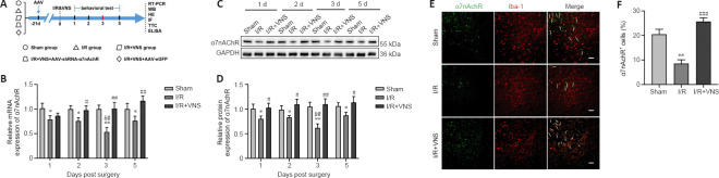
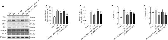
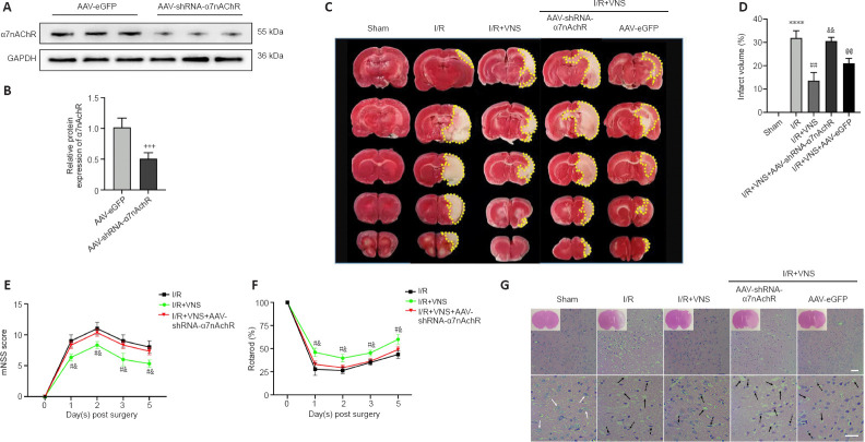
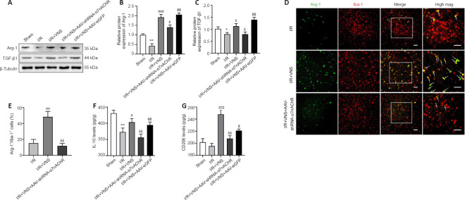
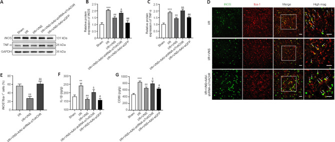
Microglia are the brain's primary innate immune cells, and they are activated and affect pro-inflammatory phenotype or regulatory phenotype after ischemic stroke. Vagus nerve stimulation was shown to activate microglial phenotypic changes and exhibit neuroprotective effects in ischemia/reperfusion injury. In this study, we established rat models of ischemic stroke by occlusion of the middle cerebral artery and performed vagus nerve stimulation 30 minutes after modeling. We found that vagus nerve stimulation caused a shift from a pro-inflammatory phenotype to a regulatory phenotype in microglia in the ischemic penumbra. Vagus nerve stimulation decreased the levels of pro-inflammatory phenotype markers inducible nitric oxide synthase and tumor necrosis factor α and increased the expression of regulatory phenotype markers arginase 1 and transforming growth factor β through activating α7 nicotinic acetylcholine receptor expression. Additionally, α7 nicotinic acetylcholine receptor blockade reduced the inhibition of Toll-like receptor 4/nuclear factor kappa-B pathway-associated proteins, including Toll-like receptor 4, myeloid differentiation factor 88, I kappa B alpha, and phosphorylated-I kappa B alpha, and also weakened the neuroprotective effects of vagus nerve stimulation in ischemic stroke. Vagus nerve stimulation inhibited Toll-like receptor 4/nuclear factor kappa-B expression through activating α7 nicotinic acetylcholine receptor and regulated microglial polarization after ischemic stroke, thereby playing a role in the treatment of ischemic stroke. Findings from this study confirm the mechanism underlying vagus nerve stimulation against ischemic stroke.
小胶质细胞是大脑主要的固有免疫细胞,在缺血性中风后会被激活并影响促炎表型或调节表型。迷走神经刺激已被证明可激活小胶质细胞的表型变化,并在缺血/再灌注损伤中发挥神经保护作用。在本研究中,我们通过大脑中动脉闭塞建立了大鼠缺血性中风模型,并在建模后30分钟进行迷走神经刺激。我们发现,迷走神经刺激导致缺血半暗带中的小胶质细胞从促炎表型转变为调节表型。迷走神经刺激通过激活α7烟碱型乙酰胆碱受体表达,降低了促炎表型标志物诱导型一氧化氮合酶和肿瘤坏死因子α的水平,并增加了调节表型标志物精氨酸酶1和转化生长因子β的表达。此外,α7烟碱型乙酰胆碱受体阻断减少了对Toll样受体4/核因子κB通路相关蛋白的抑制,包括Toll样受体4、髓样分化因子88、IκBα和磷酸化IκBα,也削弱了迷走神经刺激在缺血性中风中的神经保护作用。迷走神经刺激通过激活α7烟碱型乙酰胆碱受体抑制Toll样受体4/核因子κB表达,并在缺血性中风后调节小胶质细胞极化,从而在缺血性中风治疗中发挥作用。本研究结果证实了迷走神经刺激对抗缺血性中风的机制。