• 文献检索
  • 文档翻译
  • 深度研究
  • 学术资讯
  • Suppr Zotero 插件Zotero 插件
  • 邀请有礼
  • 套餐&价格
  • 历史记录
应用&插件
Suppr Zotero 插件Zotero 插件浏览器插件Mac 客户端Windows 客户端微信小程序
定价
高级版会员购买积分包购买API积分包
服务
文献检索文档翻译深度研究API 文档MCP 服务
关于我们
关于 Suppr公司介绍联系我们用户协议隐私条款
关注我们

Suppr 超能文献

核心技术专利:CN118964589B侵权必究
粤ICP备2023148730 号-1Suppr @ 2026

文献检索

告别复杂PubMed语法,用中文像聊天一样搜索,搜遍4000万医学文献。AI智能推荐,让科研检索更轻松。

立即免费搜索

文件翻译

保留排版,准确专业,支持PDF/Word/PPT等文件格式,支持 12+语言互译。

免费翻译文档

深度研究

AI帮你快速写综述,25分钟生成高质量综述,智能提取关键信息,辅助科研写作。

立即免费体验

基于机器学习的角膜密度测量法可疑青光眼检测。

Suspect glaucoma detection from corneal densitometry supported by machine learning.

机构信息

Department of Applied Physics, University of Zaragoza, Zaragoza, Spain.

Department of Applied Physics, University of Zaragoza, Zaragoza, Spain.

出版信息

J Optom. 2022;15 Suppl 1(Suppl 1):S12-S21. doi: 10.1016/j.optom.2022.09.002. Epub 2022 Oct 7.

DOI:10.1016/j.optom.2022.09.002
PMID:36210294
原文链接:https://pmc.ncbi.nlm.nih.gov/articles/PMC9732483/
Abstract

PURPOSE

To discriminate suspect glaucomatous from control eyes using corneal densitometry based on the statistical modeling of the pixel intensity distribution of Scheimpflug images.

METHODS

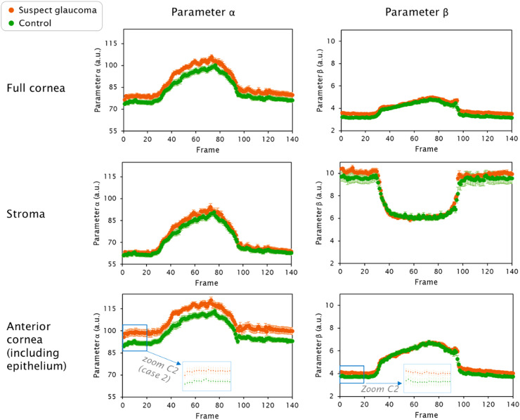
Twenty-four participants (10 suspect glaucomatous and 14 control eyes) were included in this retrospective study. Corneal biomechanics was assessed with the commercial Scheimpflug camera Corvis ST (Oculus). Sets of 140 images acquired per measurement were exported for further analysis. After corneal segmentation, pixel intensities corresponding to different corneal depths were statistically modeled for each image, from which corneal densitometry in the form of parameters α (brightness) and β (homogeneity) was derived. After data pre-processing, parameters α and β were input to six supervised machine learning algorithms that were trained, tested, and compared.

RESULTS

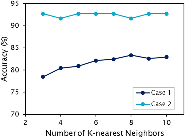
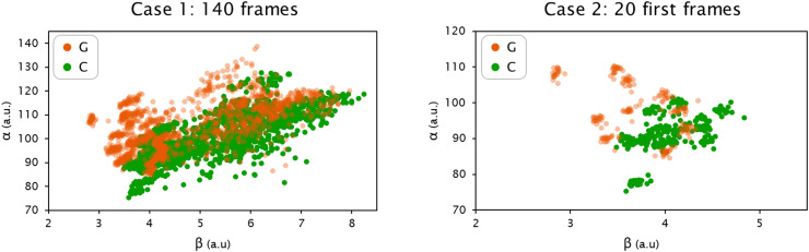
There exists a statistically significant difference in α and β parameters between suspect glaucomatous and control eyes (both, P < 0.05/N, Bonferroni). From the implemented supervised machine learning algorithms, the K-nearest neighbors (K-NN) algorithm reached 83.93% accuracy to discriminate between groups only using corneal densitometry parameters (α and β).

CONCLUSION

Densitometry of the anterior cornea including epithelium on its own has the potential to serve as a clinical tool for early glaucoma risk assessment.

摘要

目的

利用基于 Scheimpflug 图像像素强度分布的统计建模,对疑似青光眼和对照眼进行角膜密度测定,以鉴别疑似青光眼和对照眼。

方法

本回顾性研究纳入 24 名参与者(10 只疑似青光眼眼和 14 只对照眼)。采用商用 Scheimpflug 相机 Corvis ST(Oculus)评估角膜生物力学。每次测量采集 140 组图像,并对其进行进一步分析。在角膜分割后,对每张图像中不同角膜深度的像素强度进行统计建模,由此得出角膜密度参数α(亮度)和β(均匀性)。在数据预处理后,将参数α和β输入到经过训练、测试和比较的六种监督机器学习算法中。

结果

疑似青光眼眼和对照眼的α和β参数存在统计学显著差异(均 P < 0.05/N,Bonferroni)。在所实施的监督机器学习算法中,仅使用角膜密度参数(α和β),K-最近邻(K-NN)算法的分组准确率达到 83.93%。

结论

包括上皮在内的前角膜密度测定有可能成为早期青光眼风险评估的临床工具。

https://cdn.ncbi.nlm.nih.gov/pmc/blobs/0dc8/9732483/bd9893e66203/gr6.jpg
https://cdn.ncbi.nlm.nih.gov/pmc/blobs/0dc8/9732483/41c8b440dc87/gr1.jpg
https://cdn.ncbi.nlm.nih.gov/pmc/blobs/0dc8/9732483/479612b39b12/gr2.jpg
https://cdn.ncbi.nlm.nih.gov/pmc/blobs/0dc8/9732483/39d4f89f27c1/gr3.jpg
https://cdn.ncbi.nlm.nih.gov/pmc/blobs/0dc8/9732483/b1ce87b40c1e/gr4.jpg
https://cdn.ncbi.nlm.nih.gov/pmc/blobs/0dc8/9732483/3914d47efc11/gr5.jpg
https://cdn.ncbi.nlm.nih.gov/pmc/blobs/0dc8/9732483/bd9893e66203/gr6.jpg

相似文献

1
Suspect glaucoma detection from corneal densitometry supported by machine learning.基于机器学习的角膜密度测量法可疑青光眼检测。
J Optom. 2022;15 Suppl 1(Suppl 1):S12-S21. doi: 10.1016/j.optom.2022.09.002. Epub 2022 Oct 7.
2
Evaluation of corneal densitometry values with Pentacam in cases of ocular hypertension and pseudoexfoliative glaucoma.使用Pentacam评估高眼压症和假性剥脱性青光眼患者的角膜密度测量值。
Photodiagnosis Photodyn Ther. 2024 Apr;46:103988. doi: 10.1016/j.pdpdt.2024.103988. Epub 2024 Feb 16.
3
Corneal Properties of Keratoconus Based on Scheimpflug Light Intensity Distribution.基于 Scheimpflug 光强分布的圆锥角膜角膜特性。
Invest Ophthalmol Vis Sci. 2019 Jul 1;60(8):3197-3203. doi: 10.1167/iovs.19-26963.
4
Age-Related Corneal Transparency Changes Evaluated With an Alternative Method to Corneal Densitometry.用角膜密度计替代方法评估年龄相关的角膜透明度变化。
Cornea. 2021 Feb 1;40(2):215-222. doi: 10.1097/ICO.0000000000002511.
5
Detection of Subclinical Keratoconus With a Validated Alternative Method to Corneal Densitometry.利用经过验证的角膜密度测量替代方法检测亚临床圆锥角膜。
Transl Vis Sci Technol. 2021 Aug 2;10(9):32. doi: 10.1167/tvst.10.9.32.
6
Corneal biomechanical characteristics measured by the CorVis Scheimpflug technology in eyes with primary open-angle glaucoma and normal eyes.应用 Corvis ST 眼前节分析仪测量原发性开角型青光眼和正常眼的角膜生物力学特征。
Acta Ophthalmol. 2016 Aug;94(5):e317-24. doi: 10.1111/aos.12672. Epub 2015 Feb 1.
7
Corneal Stiffness Parameters Are Predictive of Structural and Functional Progression in Glaucoma Suspect Eyes.角膜硬度参数可预测青光眼疑似眼中的结构和功能进展。
Ophthalmology. 2021 Jul;128(7):993-1004. doi: 10.1016/j.ophtha.2020.11.021. Epub 2020 Nov 25.
8
Corneal Properties in Primary Open-angle Glaucoma Assessed Through Scheimpflug Corneal Topography and Densitometry.通过 Scheimpflug 角膜地形和密度测量评估原发性开角型青光眼的角膜特性。
J Glaucoma. 2021 May 1;30(5):444-450. doi: 10.1097/IJG.0000000000001770.
9
A comparison of the corneal biomechanics in pseudoexfoliation syndrome, pseudoexfoliation glaucoma, and healthy controls using Corvis® Scheimpflug Technology.应用 Corvis® Scheimpflug 技术对原发性开角型青光眼、原发性开角型青光眼伴发的假性剥脱综合征和健康对照者的角膜生物力学的比较。
Indian J Ophthalmol. 2020 May;68(5):787-792. doi: 10.4103/ijo.IJO_1550_19.
10
Corneal densitometry and biomechanical properties in patients with primary congenital glaucoma.原发性先天性青光眼患者的角膜密度测定和生物力学特性。
Can J Ophthalmol. 2021 Dec;56(6):364-370. doi: 10.1016/j.jcjo.2021.01.009. Epub 2021 Feb 10.

引用本文的文献

1
The comparison of corneal densitometry in cases with glaucoma following childhood cataract surgery and juvenile glaucoma.儿童期白内障手术后青光眼与青少年型青光眼的角膜密度比较。
Int Ophthalmol. 2024 Feb 12;44(1):64. doi: 10.1007/s10792-024-03004-0.
2
Journal of Optometry Impact Factor: it's been a long way to here.《验光杂志》影响因子:走到今天历经漫长历程。
J Optom. 2023 Jan-Mar;16(1):1-2. doi: 10.1016/j.optom.2022.12.001.

本文引用的文献

1
On the protective role of the blood vessels in glaucomatous damage: A transversal study.关于血管在青光眼性损害中的保护作用:一项横向研究。
J Optom. 2023 Jan-Mar;16(1):81-87. doi: 10.1016/j.optom.2021.10.003. Epub 2021 Dec 20.
2
Scheimpflug-Based Analysis of the Reflectivity of the Cornea in Marfan Syndrome.Scheimpflug 基于的马凡综合征角膜反射率分析。
Transl Vis Sci Technol. 2021 Aug 2;10(9):34. doi: 10.1167/tvst.10.9.34.
3
Detection of Subclinical Keratoconus With a Validated Alternative Method to Corneal Densitometry.
利用经过验证的角膜密度测量替代方法检测亚临床圆锥角膜。
Transl Vis Sci Technol. 2021 Aug 2;10(9):32. doi: 10.1167/tvst.10.9.32.
4
Corneal densitometry and biomechanical properties in patients with primary congenital glaucoma.原发性先天性青光眼患者的角膜密度测定和生物力学特性。
Can J Ophthalmol. 2021 Dec;56(6):364-370. doi: 10.1016/j.jcjo.2021.01.009. Epub 2021 Feb 10.
5
Corneal Properties in Primary Open-angle Glaucoma Assessed Through Scheimpflug Corneal Topography and Densitometry.通过 Scheimpflug 角膜地形和密度测量评估原发性开角型青光眼的角膜特性。
J Glaucoma. 2021 May 1;30(5):444-450. doi: 10.1097/IJG.0000000000001770.
6
Supervised machine learning tools: a tutorial for clinicians.监督机器学习工具:临床医生教程。
J Neural Eng. 2020 Nov 19;17(6). doi: 10.1088/1741-2552/abbff2.
7
Age-Related Corneal Transparency Changes Evaluated With an Alternative Method to Corneal Densitometry.用角膜密度计替代方法评估年龄相关的角膜透明度变化。
Cornea. 2021 Feb 1;40(2):215-222. doi: 10.1097/ICO.0000000000002511.
8
Keratoconus Detection Based on a Single Scheimpflug Image.基于单张眼前节光学生物测量仪图像的圆锥角膜检测
Transl Vis Sci Technol. 2020 Jun 26;9(7):36. doi: 10.1167/tvst.9.7.36. eCollection 2020 Jun.
9
Corneal densitometry and topography in patients with primary congenital glaucoma.原发性先天性青光眼患者的角膜密度测量与地形图分析
J Fr Ophtalmol. 2020 Oct;43(8):697-703. doi: 10.1016/j.jfo.2019.11.035. Epub 2020 Aug 10.
10
Corneal tissue properties following scleral lens wear using Scheimpflug imaging.使用Scheimpflug成像技术观察巩膜镜佩戴后角膜组织特性
Ophthalmic Physiol Opt. 2020 Sep;40(5):595-606. doi: 10.1111/opo.12710. Epub 2020 Jul 23.