• 文献检索
  • 文档翻译
  • 深度研究
  • 学术资讯
  • Suppr Zotero 插件Zotero 插件
  • 邀请有礼
  • 套餐&价格
  • 历史记录
应用&插件
Suppr Zotero 插件Zotero 插件浏览器插件Mac 客户端Windows 客户端微信小程序
定价
高级版会员购买积分包购买API积分包
服务
文献检索文档翻译深度研究API 文档MCP 服务
关于我们
关于 Suppr公司介绍联系我们用户协议隐私条款
关注我们

Suppr 超能文献

核心技术专利:CN118964589B侵权必究
粤ICP备2023148730 号-1Suppr @ 2026

文献检索

告别复杂PubMed语法,用中文像聊天一样搜索,搜遍4000万医学文献。AI智能推荐,让科研检索更轻松。

立即免费搜索

文件翻译

保留排版,准确专业,支持PDF/Word/PPT等文件格式,支持 12+语言互译。

免费翻译文档

深度研究

AI帮你快速写综述,25分钟生成高质量综述,智能提取关键信息,辅助科研写作。

立即免费体验

使用动态超广角红外共焦扫描激光检眼镜对玻璃体混浊进行可视化和分级:一项初步研究。

Visualization and Grading of Vitreous Floaters Using Dynamic Ultra-Widefield Infrared Confocal Scanning Laser Ophthalmoscopy: A Pilot Study.

作者信息

Garcia-Aguirre Gerardo, Henaine-Berra Andree, Salcedo-Villanueva Guillermo

机构信息

Retina Department, Asociación para Evitar la Ceguera en México I.A.P., Mexico City 04030, Mexico.

School of Medicine and Health Sciences, Tecnologico de Monterrey, Mexico City 14380, Mexico.

出版信息

J Clin Med. 2022 Sep 20;11(19):5502. doi: 10.3390/jcm11195502.

DOI:10.3390/jcm11195502
PMID:36233369
原文链接:https://pmc.ncbi.nlm.nih.gov/articles/PMC9571201/
Abstract

PURPOSE

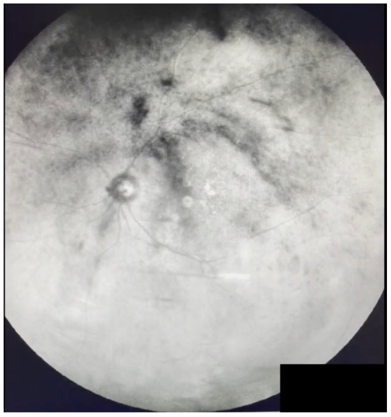
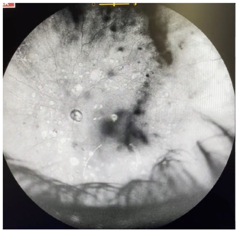
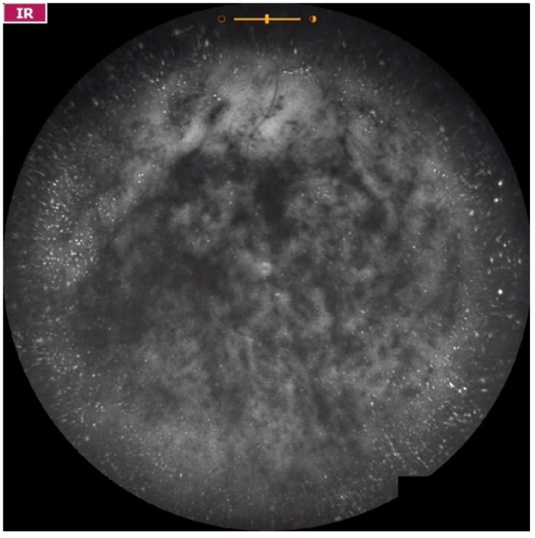
To describe the appearance of vitreous opacities using dynamic ultra-widefield infrared confocal scanning laser ophthalmoscopy (IRcSLO).

DESIGN

Retrospective case series.

METHODS

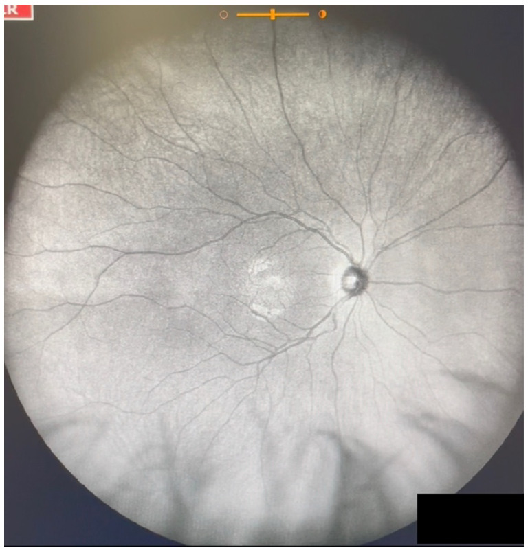
Eyes of patients complaining of myodesopsia were analyzed using dynamic ultra-widefield IRcSLO imaging (Nidek Mirante, Nidek Co., Ltd., Gamagori, Japan), and classified according to a vitreous opacity severity scale.

RESULTS

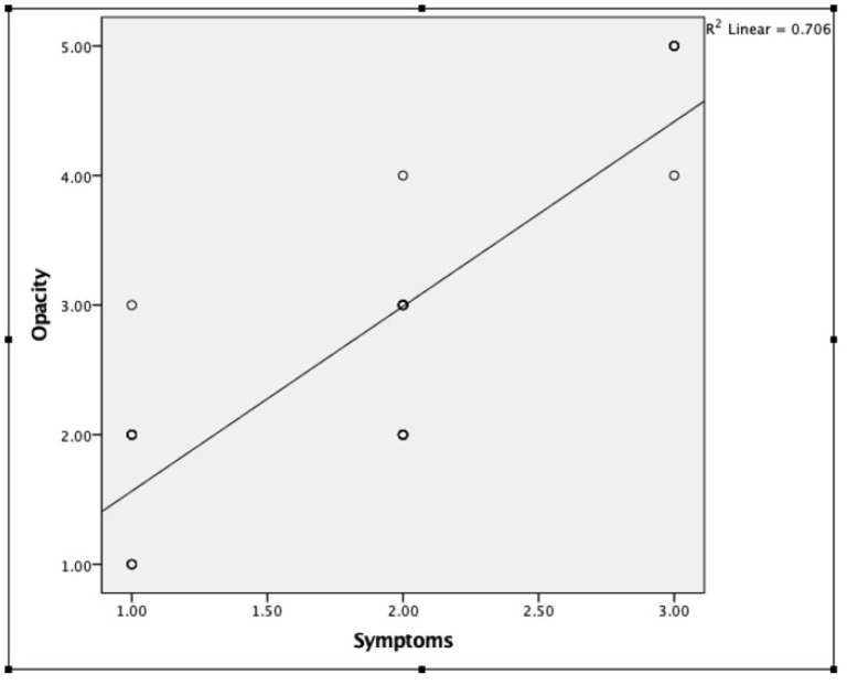
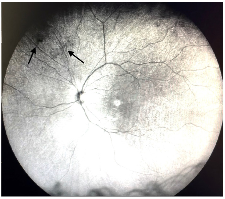
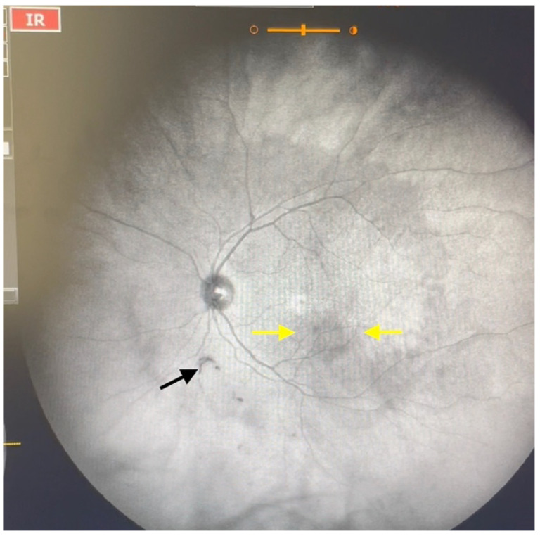
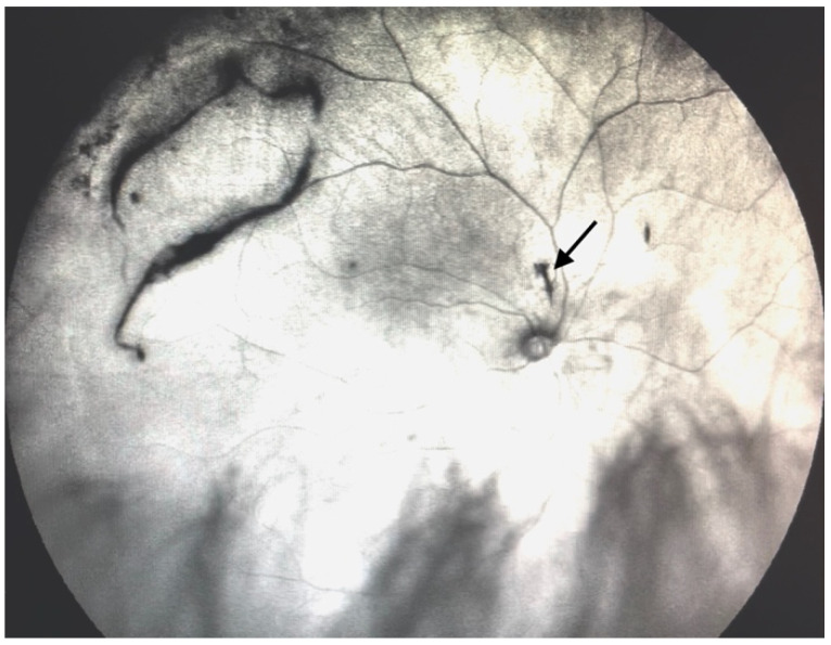
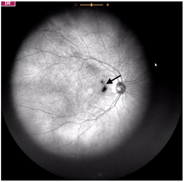
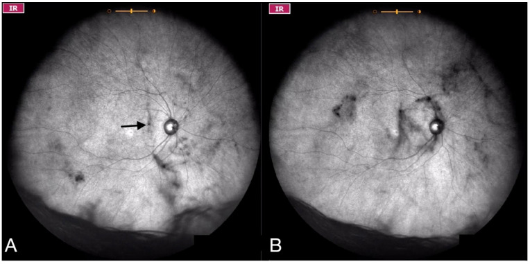
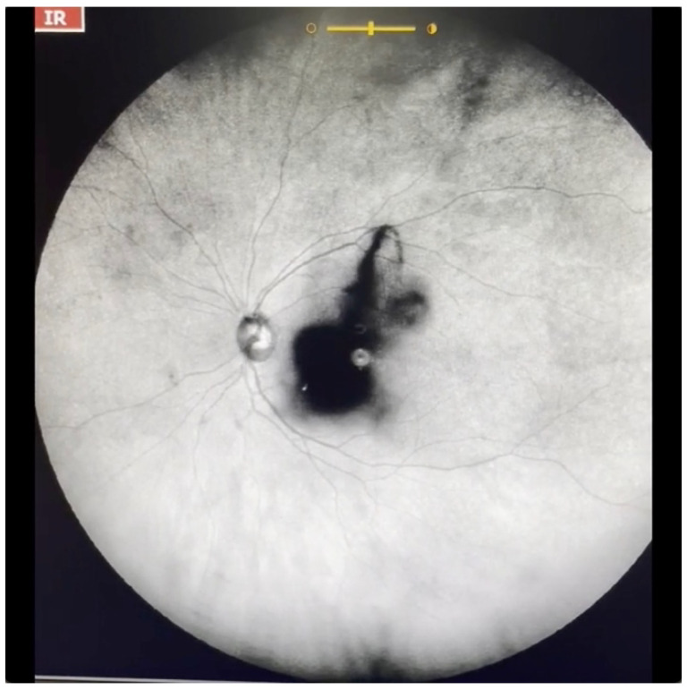
Thirty eyes of 21 patients were included in this study. The average age was 56 years. Symptom duration ranged from 1 to more than 365 days. The most common cause of vitreous floaters was posterior vitreous detachment (63.3%), followed by vitreous syneresis (23.3%), asteroid hyalosis (10%) and vitreous hemorrhage (3.3%). Opacities were classified as Grade 1 in three eyes (10%), Grade 2 in 10 eyes (33.3%), Grade 3 in 11 eyes (36.6%), Grade 4 in two eyes (6.6%) and Grade 5 in four eyes (13.3%). Patients with Grade 1 opacities were younger than patients with opacities Grade 2 or greater. A visible Weiss ring could be identified in 0% of eyes with Grade 1 opacities, 40% of eyes with Grade 2 opacities, 100% of eyes with Grade 3 opacities, and 100% of eyes with Grade 4 opacities. In patients with Grade 5 opacities, a Weiss ring could not be identified.

CONCLUSION

Dynamic ultra-widefield IRcSLO imaging is a useful tool to evaluate patients with vitreous floaters. It allows for accurate visualization of the number, density, and behavior of the shadows that vitreous opacities project over a very wide area of the retina, which has a positive correlation with patient perception of floaters.

摘要

目的

使用动态超广角红外共焦扫描激光检眼镜(IRcSLO)描述玻璃体混浊的表现。

设计

回顾性病例系列。

方法

使用动态超广角IRcSLO成像(日本蒲郡市尼德克公司的尼德克Mirante)对主诉有飞蚊症的患者眼睛进行分析,并根据玻璃体混浊严重程度量表进行分类。

结果

本研究纳入了21例患者的30只眼睛。平均年龄为56岁。症状持续时间为1天至超过365天。玻璃体混浊最常见的原因是玻璃体后脱离(63.3%),其次是玻璃体液化(23.3%)、星状玻璃体变性(10%)和玻璃体积血(3.3%)。混浊被分类为1级的有3只眼(10%),2级的有10只眼(33.3%),3级的有11只眼(36.6%),4级的有2只眼(6.6%),5级的有4只眼(13.3%)。1级混浊患者比2级及以上混浊患者年轻。1级混浊眼中0%能识别出可见的魏氏环,2级混浊眼中40%能识别出,3级混浊眼中100%能识别出,4级混浊眼中100%能识别出。5级混浊患者中无法识别出魏氏环。

结论

动态超广角IRcSLO成像对于评估有玻璃体混浊的患者是一种有用的工具。它能够准确显示玻璃体混浊在视网膜非常大的区域投射的阴影的数量、密度和动态,这与患者对飞蚊症的感知呈正相关。

https://cdn.ncbi.nlm.nih.gov/pmc/blobs/c195/9571201/db686905c75f/jcm-11-05502-g011.jpg
https://cdn.ncbi.nlm.nih.gov/pmc/blobs/c195/9571201/f31748fbe9a3/jcm-11-05502-g001.jpg
https://cdn.ncbi.nlm.nih.gov/pmc/blobs/c195/9571201/ab7450309451/jcm-11-05502-g002.jpg
https://cdn.ncbi.nlm.nih.gov/pmc/blobs/c195/9571201/3c6de8263edb/jcm-11-05502-g003.jpg
https://cdn.ncbi.nlm.nih.gov/pmc/blobs/c195/9571201/30814310ee3c/jcm-11-05502-g004.jpg
https://cdn.ncbi.nlm.nih.gov/pmc/blobs/c195/9571201/88a087655da8/jcm-11-05502-g005.jpg
https://cdn.ncbi.nlm.nih.gov/pmc/blobs/c195/9571201/e6b5ca5887b7/jcm-11-05502-g006.jpg
https://cdn.ncbi.nlm.nih.gov/pmc/blobs/c195/9571201/f0f6708053c0/jcm-11-05502-g007.jpg
https://cdn.ncbi.nlm.nih.gov/pmc/blobs/c195/9571201/ba521f3c92d4/jcm-11-05502-g008.jpg
https://cdn.ncbi.nlm.nih.gov/pmc/blobs/c195/9571201/58a576eaf649/jcm-11-05502-g009.jpg
https://cdn.ncbi.nlm.nih.gov/pmc/blobs/c195/9571201/f6ecb31c4e89/jcm-11-05502-g010.jpg
https://cdn.ncbi.nlm.nih.gov/pmc/blobs/c195/9571201/db686905c75f/jcm-11-05502-g011.jpg

相似文献

1
Visualization and Grading of Vitreous Floaters Using Dynamic Ultra-Widefield Infrared Confocal Scanning Laser Ophthalmoscopy: A Pilot Study.使用动态超广角红外共焦扫描激光检眼镜对玻璃体混浊进行可视化和分级:一项初步研究。
J Clin Med. 2022 Sep 20;11(19):5502. doi: 10.3390/jcm11195502.
2
New Terminology and Methodology for the Assessment of the Vitreous, Its Floaters and Opacities, and Their Effect on Vision: Standardized and Kinetic Anatomical and Functional Testing of Vitreous Floaters and Opacities (SK VFO Test).评估玻璃体、其飞蚊症和混浊物及其对视力影响的新术语和方法:玻璃体飞蚊症和混浊物的标准化动态解剖学和功能测试(SK VFO测试)
Ophthalmic Surg Lasers Imaging Retina. 2023 May;54(5):306-315. doi: 10.3928/23258160-20230412-02. Epub 2023 May 1.
3
Vision degrading myodesopsia assessed with optos ultra-widefield scanning laser ophthalmoscope.采用光学生物测量仪超广角扫描激光检眼镜评估进行性视力丧失性眼球震颤。
BMC Ophthalmol. 2023 Oct 20;23(1):425. doi: 10.1186/s12886-023-03166-y.
4
[Evaluation of YAG-laser vitreolysis effectiveness based on quantitative characterization of vitreous floaters].基于玻璃体混浊定量表征评估YAG激光玻璃体溶解术的有效性
Vestn Oftalmol. 2018;134(1):56-62. doi: 10.17116/oftalma2018134156-62.
5
Practical applications of vitreous imaging for the treatment of vitreous opacities with YAG vitreolysis.玻璃体成像在YAG玻璃体溶解术治疗玻璃体混浊中的实际应用
Int Ophthalmol. 2023 Oct;43(10):3587-3594. doi: 10.1007/s10792-023-02765-4. Epub 2023 Jul 4.
6
Dynamic Infrared Imaging of Vitreous Floaters.玻璃体混浊的动态红外成像
Clin Ophthalmol. 2023 Apr 17;17:1169-1177. doi: 10.2147/OPTH.S399684. eCollection 2023.
7
Acute symptomatic vitreous floaters assessed with ultra-wide field scanning laser ophthalmoscopy and spectral domain optical coherence tomography.应用超广角扫描激光检眼镜和谱域光相干断层扫描评估急性症状性玻璃体浮游物。
Sci Rep. 2021 Apr 26;11(1):8930. doi: 10.1038/s41598-021-88371-9.
8
Predictive value of floaters in the diagnosis of posterior vitreous detachment.飞蚊症在诊断玻璃体后脱离中的预测价值。
Am J Ophthalmol. 1998 Jan;125(1):113-5. doi: 10.1016/s0002-9394(99)80250-5.
9
THE WAY PATIENTS SEE FLOATERS: Widefield Dynamic Scanning Laser Ophthalmoscopy Imaging of Vitreous Abnormalities.患者眼中的飞蚊症:玻璃体异常的广角动态扫描激光眼底镜成像
Retina. 2023 Aug 1;43(8):1240-1245. doi: 10.1097/IAE.0000000000003786.
10
Ultra-widefield fundus imaging in gas-filled eyes after vitrectomy.玻璃体切除术后充气眼的超广角眼底成像
BMC Ophthalmol. 2017 Jul 3;17(1):114. doi: 10.1186/s12886-017-0510-7.

引用本文的文献

1
Infrared Video Imaging for the Identification and Quantification of Macula-Involving Symptomatic Vitreous Opacities.用于识别和量化累及黄斑的有症状玻璃体混浊的红外视频成像
J Vitreoretin Dis. 2025 Jan 9:24741264241310219. doi: 10.1177/24741264241310219.
2
Management of Visually Significant Vitreous Opacities.显著玻璃体混浊的处理。
Mo Med. 2024 Sep-Oct;121(5):391-394.
3
Forward Light Scattering of the Vitreous Gel After Enzymatic Aging: An In Vitro Model to Study Vitreous Opacification.酶老化后玻璃体凝胶的前向光散射:研究玻璃体混浊的体外模型。

本文引用的文献

1
Volumetric Optical Imaging and Quantitative Analysis of Age-Related Changes in Anterior Human Vitreous.容积光学成像及人眼前房玻璃体年龄相关性改变的定量分析。
Invest Ophthalmol Vis Sci. 2021 Apr 1;62(4):31. doi: 10.1167/iovs.62.4.31.
2
Acute symptomatic vitreous floaters assessed with ultra-wide field scanning laser ophthalmoscopy and spectral domain optical coherence tomography.应用超广角扫描激光检眼镜和谱域光相干断层扫描评估急性症状性玻璃体浮游物。
Sci Rep. 2021 Apr 26;11(1):8930. doi: 10.1038/s41598-021-88371-9.
3
Vitreous Structure and Visual Function in Myopic Vitreopathy Causing Vision-Degrading Myodesopsia.
Invest Ophthalmol Vis Sci. 2024 Mar 5;65(3):36. doi: 10.1167/iovs.65.3.36.
4
Patients with floaters: Answers from virtual assistants and large language models.患有飞蚊症的患者:来自虚拟助手和大语言模型的回答。
Digit Health. 2024 Feb 14;10:20552076241229933. doi: 10.1177/20552076241229933. eCollection 2024 Jan-Dec.
5
Visualization and Identification of Silicone Oil Emulsification Using Dynamic Infrared Confocal Scanning Laser Ophthalmoscopy.使用动态红外共焦扫描激光眼科显微镜对硅油乳化进行可视化和识别
Case Rep Ophthalmol. 2024 Jan 9;15(1):36-40. doi: 10.1159/000535746. eCollection 2024 Jan-Dec.
6
Vision degrading myodesopsia assessed with optos ultra-widefield scanning laser ophthalmoscope.采用光学生物测量仪超广角扫描激光检眼镜评估进行性视力丧失性眼球震颤。
BMC Ophthalmol. 2023 Oct 20;23(1):425. doi: 10.1186/s12886-023-03166-y.
7
Spotlight on Lattice Degeneration Imaging Techniques.聚焦于格子样变性成像技术。
Clin Ophthalmol. 2023 Aug 16;17:2383-2395. doi: 10.2147/OPTH.S405200. eCollection 2023.
8
Dynamic Infrared Imaging of Vitreous Floaters.玻璃体混浊的动态红外成像
Clin Ophthalmol. 2023 Apr 17;17:1169-1177. doi: 10.2147/OPTH.S399684. eCollection 2023.
9
A New Pharmacological Vitreolysis through the Supplement of Mixed Fruit Enzymes for Patients with Ocular Floaters or Vitreous Hemorrhage-Induced Floaters.通过补充混合水果酶对飞蚊症或玻璃体出血引起的飞蚊症患者进行新型药理玻璃体溶解术。
J Clin Med. 2022 Nov 13;11(22):6710. doi: 10.3390/jcm11226710.
近视性玻璃体病变导致视力下降性飞蚊症的玻璃体结构与视觉功能
Am J Ophthalmol. 2021 Apr;224:246-253. doi: 10.1016/j.ajo.2020.09.017. Epub 2020 Sep 17.
4
Reading Speed as an Objective Measure of Improvement Following Vitrectomy for Symptomatic Vitreous Opacities.阅读速度作为玻璃体切割术后改善症状性玻璃体混浊的客观衡量指标。
Ophthalmic Surg Lasers Imaging Retina. 2020 Aug 1;51(8):456-466. doi: 10.3928/23258160-20200804-06.
5
Standard 6-mm Compared with Widefield 16.5-mm OCT for Staging of Posterior Vitreous Detachment.6 毫米标准与 16.5 毫米广角 OCT 在后脱离中的分期比较。
Ophthalmol Retina. 2020 Nov;4(11):1093-1102. doi: 10.1016/j.oret.2020.05.006. Epub 2020 May 19.
6
A Proposed Method to Quantify Vitreous Hemorrhage by Ultrasound.
Clin Ophthalmol. 2019 Dec 2;13:2377-2384. doi: 10.2147/OPTH.S229857. eCollection 2019.
7
Assessment of Vitreous Structure and Visual Function after Neodymium:Yttrium-Aluminum-Garnet Laser Vitreolysis.钕:钇-铝-石榴石激光玻璃体溶解术后玻璃体结构和视觉功能评估。
Ophthalmology. 2019 Nov;126(11):1517-1526. doi: 10.1016/j.ophtha.2019.06.021. Epub 2019 Jun 22.
8
Nd:YAG Laser Vitreolysis for Symptomatic Vitreous Floaters: Application of Infrared Fundus Photography in Assessing the Treatment Efficacy.钕钇铝石榴石激光玻璃体溶解术治疗有症状的玻璃体混浊:红外眼底摄影在评估治疗效果中的应用
J Ophthalmol. 2019 Jan 27;2019:8956952. doi: 10.1155/2019/8956952. eCollection 2019.
9
Posterior Vitreous Detachment as Observed by Wide-Angle OCT Imaging.宽角 OCT 成像观察到的后玻璃体脱离。
Ophthalmology. 2018 Sep;125(9):1372-1383. doi: 10.1016/j.ophtha.2018.02.039. Epub 2018 Apr 7.
10
[Evaluation of YAG-laser vitreolysis effectiveness based on quantitative characterization of vitreous floaters].基于玻璃体混浊定量表征评估YAG激光玻璃体溶解术的有效性
Vestn Oftalmol. 2018;134(1):56-62. doi: 10.17116/oftalma2018134156-62.