Pellegrini Evelin, Desando Giovanna, Petretta Mauro, Cellamare Antonella, Cristalli Camilla, Pasello Michela, Manara Maria Cristina, Grigolo Brunella, Scotlandi Katia
Laboratory of Experimental Oncology, IRCCS Istituto Ortopedico Rizzoli, Via di Barbiano 1/10, 40136 Bologna, Italy.
Laboratory RAMSES, IRCCS Istituto Ortopedico Rizzoli, Via di Barbiano 1/10, 40136 Bologna, Italy.
Polymers (Basel). 2022 Sep 28;14(19):4070. doi: 10.3390/polym14194070.
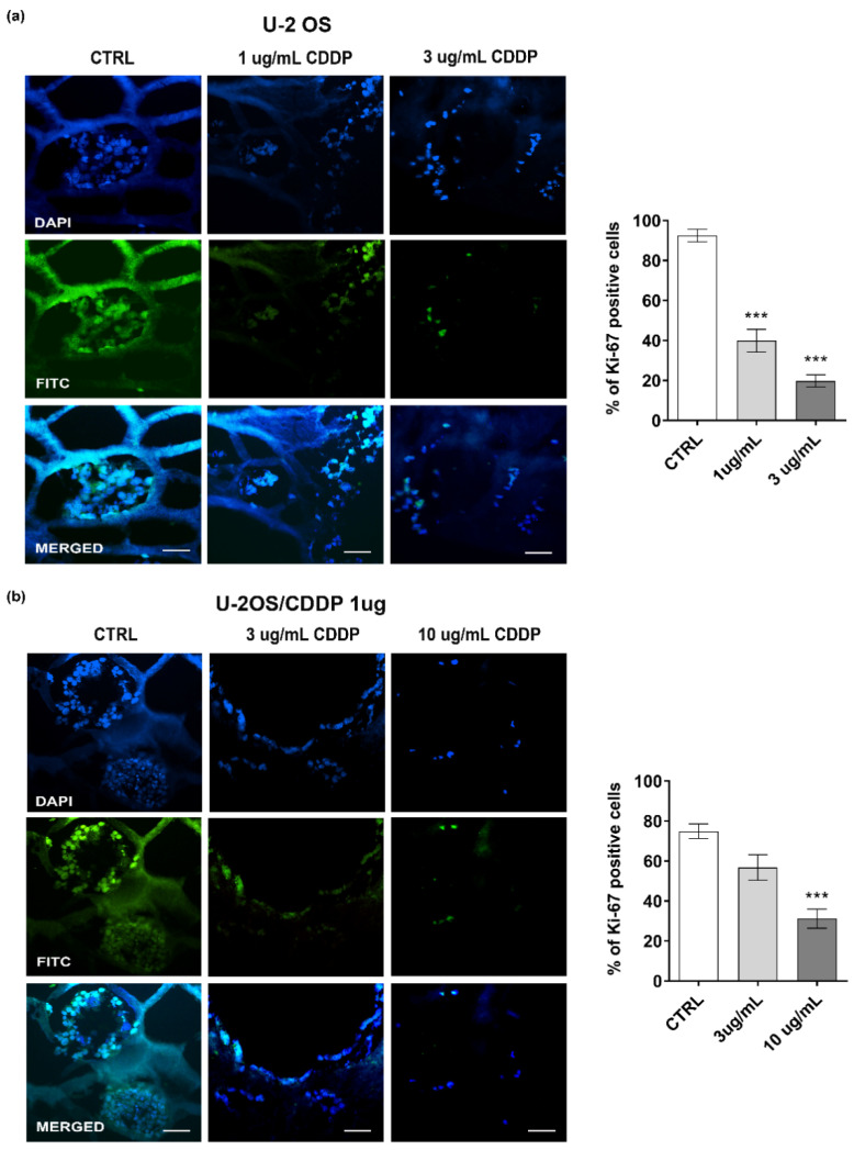
The biological and therapeutic limits of traditional 2D culture models, which only partially mimic the complexity of cancer, have recently emerged. In this study, we used a 3D bioprinting platform to process a collagen-based hydrogel with embedded osteosarcoma (OS) cells. The human OS U-2 OS cell line and its resistant variant (U-2OS/CDDP 1 μg) were considered. The fabrication parameters were optimized to obtain 3D printed constructs with overall morphology and internal microarchitecture that accurately match the theoretical design, in a reproducible and stable process. The biocompatibility of the 3D bioprinting process and the chosen collagen bioink in supporting OS cell viability and metabolism was confirmed through multiple assays at short- (day 3) and long- (day 10) term follow-ups. In addition, we tested how the 3D collagen-based bioink affects the tumor cell invasive capabilities and chemosensitivity to cisplatin (CDDP). Overall, we developed a new 3D culture model of OS cells that is easy to set up, allows reproducible results, and better mirrors malignant features of OS than flat conditions, thus representing a promising tool for drug screening and OS cell biology research.
传统二维培养模型的生物学和治疗局限性最近已显现出来,该模型仅部分模拟了癌症的复杂性。在本研究中,我们使用了一个3D生物打印平台来处理嵌入骨肉瘤(OS)细胞的基于胶原蛋白的水凝胶。我们考虑了人OS U-2 OS细胞系及其耐药变体(U-2OS/CDDP 1 μg)。优化了制造参数,以在可重复且稳定的过程中获得整体形态和内部微结构与理论设计精确匹配的3D打印构建体。通过在短期(第3天)和长期(第10天)随访中的多项测定,证实了3D生物打印过程和所选胶原蛋白生物墨水在支持OS细胞活力和代谢方面的生物相容性。此外,我们测试了基于3D胶原蛋白的生物墨水如何影响肿瘤细胞的侵袭能力和对顺铂(CDDP)的化学敏感性。总体而言,我们开发了一种新的OS细胞3D培养模型,该模型易于建立,可产生可重复的结果,并且比平面条件更好地反映了OS的恶性特征,因此是药物筛选和OS细胞生物学研究的有前途的工具。