Bioinformatics Institute (BII), Agency for Science, Technology and Research (A*STAR), Singapore 138671, Singapore.
Department of Biological Sciences, National University of Singapore, Singapore 117543, Singapore.
J Mol Cell Biol. 2023 Feb 7;14(9). doi: 10.1093/jmcb/mjac058.
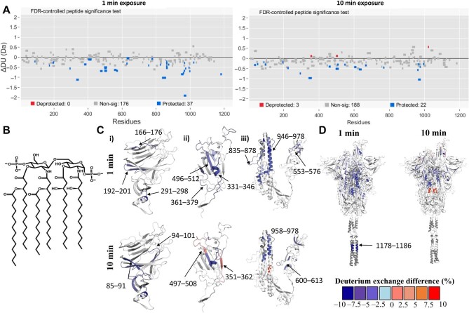
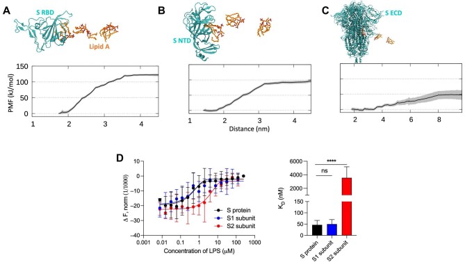
Accumulating evidence indicates a potential role for bacterial lipopolysaccharide (LPS) in the overactivation of the immune response during SARS-CoV-2 infection. LPS is recognized by Toll-like receptor 4, mediating proinflammatory effects. We previously reported that LPS directly interacts with SARS-CoV-2 spike (S) protein and enhances proinflammatory activities. Using native gel electrophoresis and hydrogen-deuterium exchange mass spectrometry, we showed that LPS binds to multiple hydrophobic pockets spanning both the S1 and S2 subunits of the S protein. Molecular simulations validated by a microscale thermophoresis binding assay revealed that LPS binds to the S2 pocket with a lower affinity compared to S1, suggesting a role as an intermediate in LPS transfer. Congruently, nuclear factor-kappa B (NF-κB) activation in monocytic THP-1 cells is strongly boosted by S2. Using NF-κB reporter mice followed by bioimaging, a boosting effect was observed for both S1 and S2, with the former potentially facilitated by proteolysis. The Omicron S variant binds to LPS, but with reduced affinity and LPS boosting in vitro and in vivo. Taken together, the data provide a molecular mechanism by which S protein augments LPS-mediated hyperinflammation.
越来越多的证据表明,细菌脂多糖 (LPS) 在 SARS-CoV-2 感染期间过度激活免疫反应中可能发挥作用。LPS 通过 Toll 样受体 4 被识别,介导促炎作用。我们之前报道过 LPS 可直接与 SARS-CoV-2 刺突 (S) 蛋白相互作用,并增强促炎活性。通过天然凝胶电泳和氢氘交换质谱,我们表明 LPS 结合到 S 蛋白的 S1 和 S2 亚基上跨越多个疏水性口袋。通过微量热泳动结合测定验证的分子模拟表明,LPS 与 S2 口袋的结合亲和力低于 S1,表明 LPS 作为 LPS 转移的中间物发挥作用。一致地,单核细胞 THP-1 细胞中的核因子-κB (NF-κB) 激活被 S2 强烈增强。使用 NF-κB 报告基因小鼠进行生物成像后,观察到 S1 和 S2 均具有增强作用,前者可能通过蛋白水解作用得到促进。Omicron S 变体与 LPS 结合,但亲和力降低,体外和体内的 LPS 增强作用降低。总之,这些数据提供了一种分子机制,解释了 S 蛋白如何增强 LPS 介导的过度炎症反应。