Wasserman Aaron H, Huang Amanda R, Lewis-Israeli Yonatan R, Dooley McKenna D, Mitchell Allison L, Venkatesan Manigandan, Aguirre Aitor
Division of Developmental and Stem Cell Biology, Institute for Quantitative Health Science and Engineering (IQ), Michigan State University, East Lansing, MI, United States.
Department of Biomedical Engineering, College of Engineering, Michigan State University, East Lansing, MI, United States.
Front Cell Dev Biol. 2022 Sep 30;10:985298. doi: 10.3389/fcell.2022.985298. eCollection 2022.
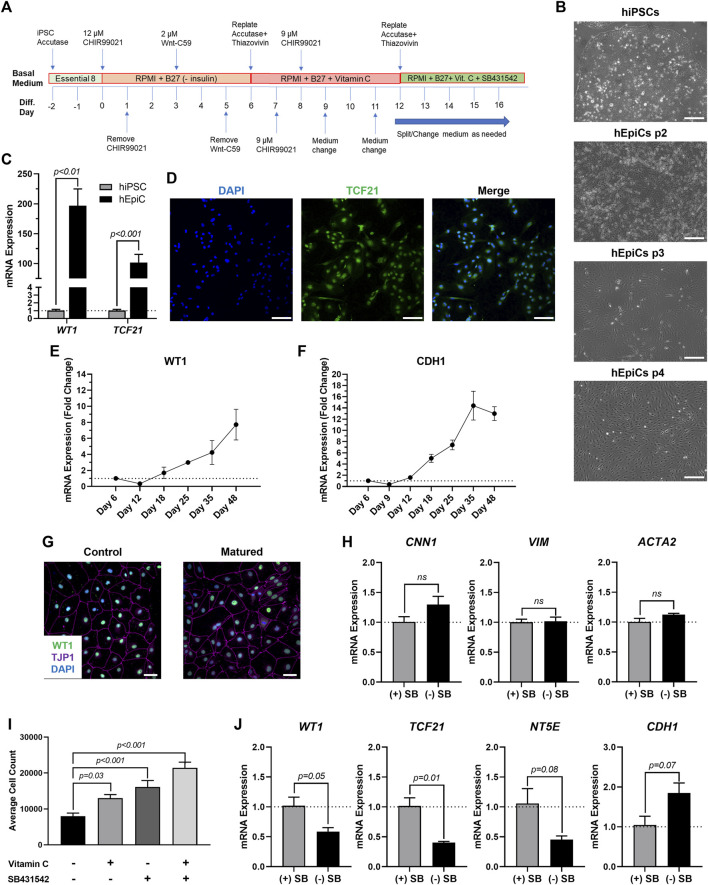
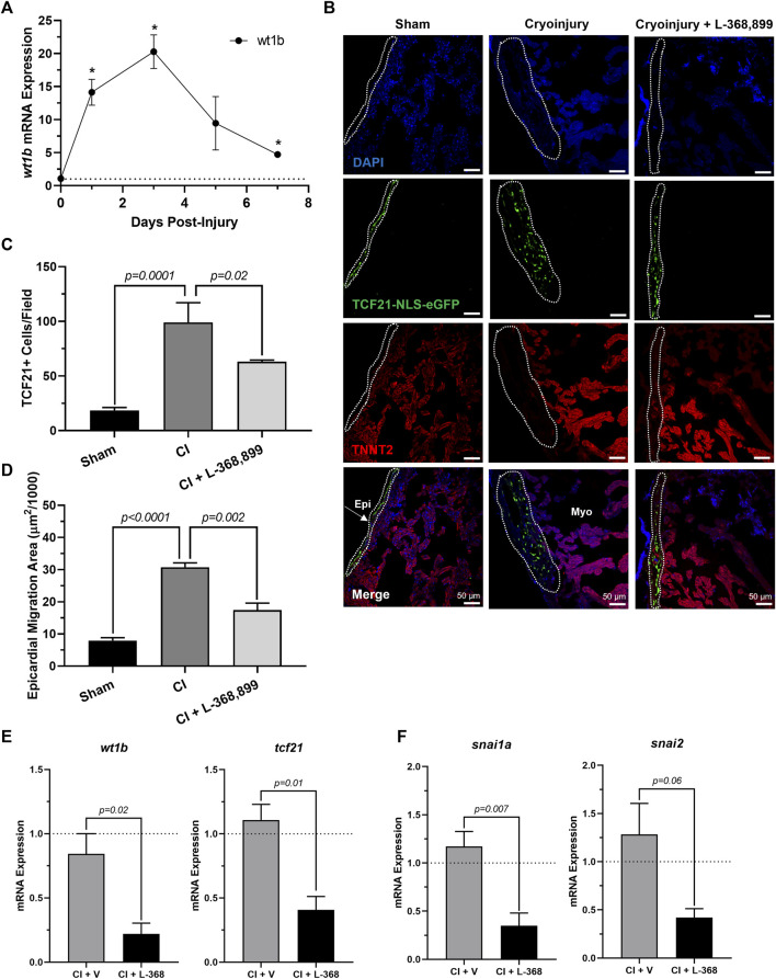
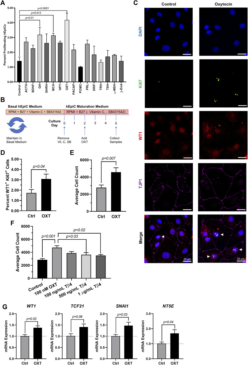
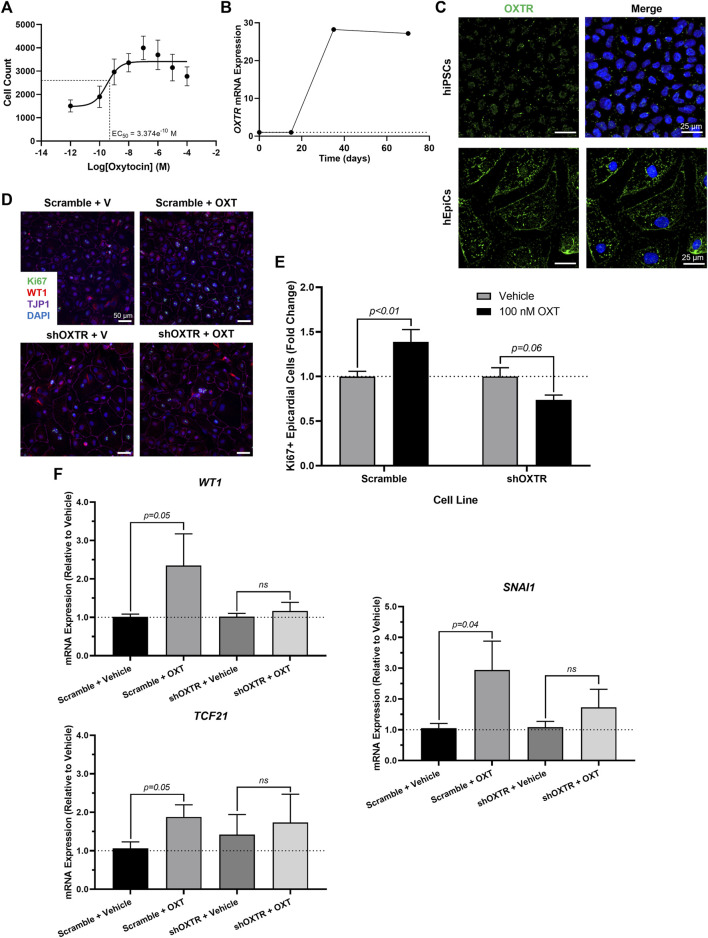
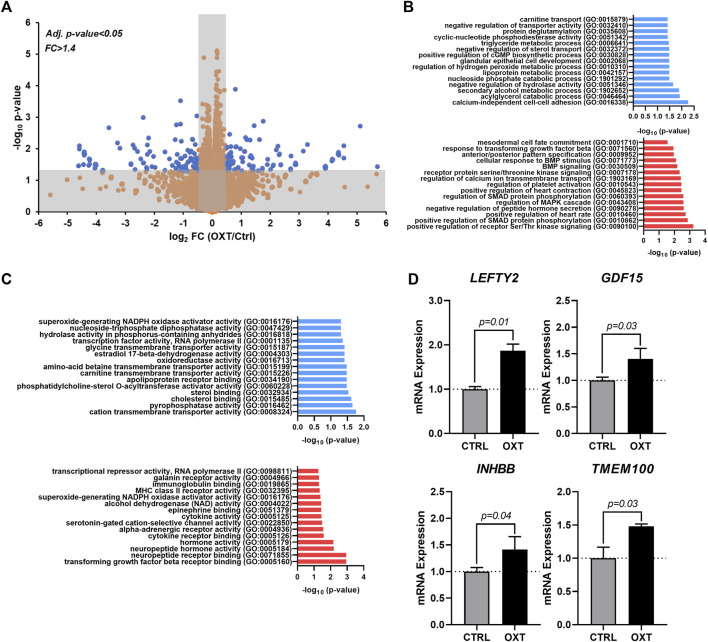
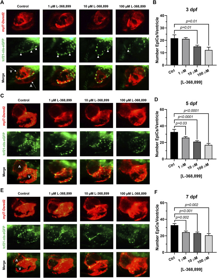
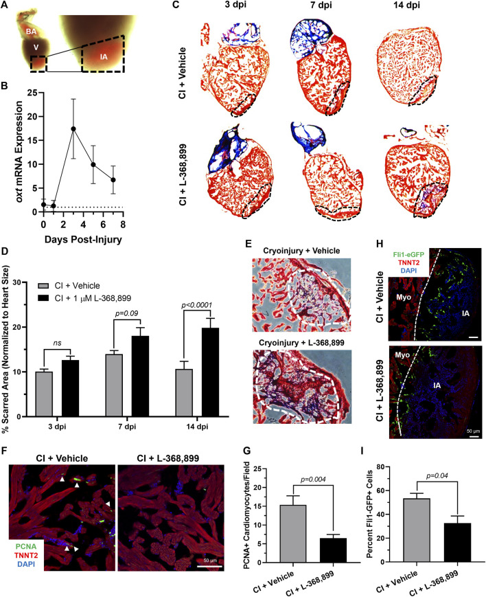
Cardiovascular disease (CVD) is one of the leading causes of mortality worldwide, and frequently leads to massive heart injury and the loss of billions of cardiac muscle cells and associated vasculature. Critical work in the last 2 decades demonstrated that these lost cells can be partially regenerated by the epicardium, the outermost mesothelial layer of the heart, in a process that highly recapitulates its role in heart development. Upon cardiac injury, mature epicardial cells activate and undergo an epithelial-mesenchymal transition (EMT) to form epicardium-derived progenitor cells (EpiPCs), multipotent progenitors that can differentiate into several important cardiac lineages, including cardiomyocytes and vascular cells. In mammals, this process alone is insufficient for significant regeneration, but it might be possible to prime it by administering specific reprogramming factors, leading to enhanced EpiPC function. Here, we show that oxytocin (OXT), a hypothalamic neuroendocrine peptide, induces epicardial cell proliferation, EMT, and transcriptional activity in a model of human induced pluripotent stem cell (hiPSC)-derived epicardial cells. In addition, we demonstrate that OXT is produced after cardiac cryoinjury in zebrafish, and that it elicits significant epicardial activation promoting heart regeneration. Oxytocin signaling is also critical for proper epicardium development in zebrafish embryos. The above processes are significantly impaired when OXT signaling is inhibited chemically or genetically through RNA interference. RNA sequencing data suggests that the transforming growth factor beta (TGF-β) pathway is the primary mediator of OXT-induced epicardial activation. Our research reveals for the first time an evolutionary conserved brain-controlled mechanism inducing cellular reprogramming and regeneration of the injured mammalian and zebrafish heart, a finding that could contribute to translational advances for the treatment of cardiac injuries.
心血管疾病(CVD)是全球主要的死亡原因之一,常导致严重的心脏损伤以及数十亿心肌细胞和相关脉管系统的丧失。过去20年的重要研究表明,这些丢失的细胞可由心脏最外层的间皮细胞层——心外膜部分再生,这一过程高度重现了其在心脏发育中的作用。心脏损伤时,成熟的心外膜细胞激活并经历上皮-间充质转化(EMT),形成心外膜衍生祖细胞(EpiPCs),即多能祖细胞,可分化为几种重要的心脏谱系,包括心肌细胞和血管细胞。在哺乳动物中,仅这一过程不足以实现显著的再生,但通过施用特定的重编程因子可能会启动这一过程,从而增强EpiPC的功能。在此,我们表明,下丘脑神经内分泌肽催产素(OXT)在人诱导多能干细胞(hiPSC)衍生的心外膜细胞模型中可诱导心外膜细胞增殖、EMT和转录活性。此外,我们证明斑马鱼心脏冷冻损伤后会产生OXT,且它能引发显著的心外膜激活,促进心脏再生。催产素信号传导对斑马鱼胚胎心外膜的正常发育也至关重要。当通过化学方法或RNA干扰进行基因抑制时,上述过程会受到显著损害。RNA测序数据表明,转化生长因子β(TGF-β)途径是OXT诱导心外膜激活的主要介导因子。我们的研究首次揭示了一种进化保守的脑控机制,可诱导受损哺乳动物和斑马鱼心脏的细胞重编程和再生,这一发现可能有助于推动心脏损伤治疗的转化进展。