Department of Drug Science, Laboratory of Cell Physiology and Molecular Neuroscience, N.I.S. Centre, University of Torino, Corso Raffaello 30, 10125, Turin, Italy.
Department of Neuroscience, University of Torino, 10125, Turin, Italy.
Pflugers Arch. 2023 Feb;475(2):181-202. doi: 10.1007/s00424-022-02761-0. Epub 2022 Oct 19.
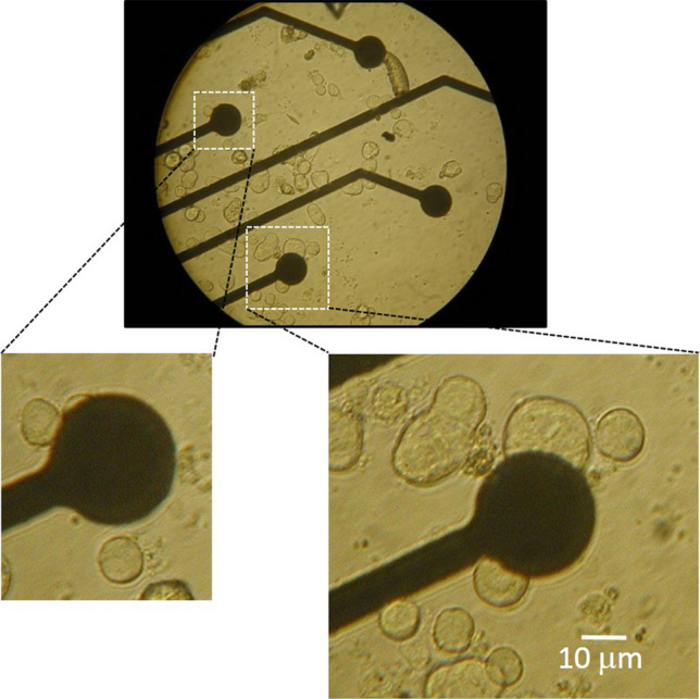
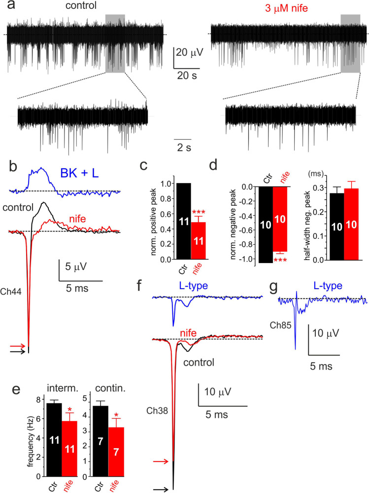
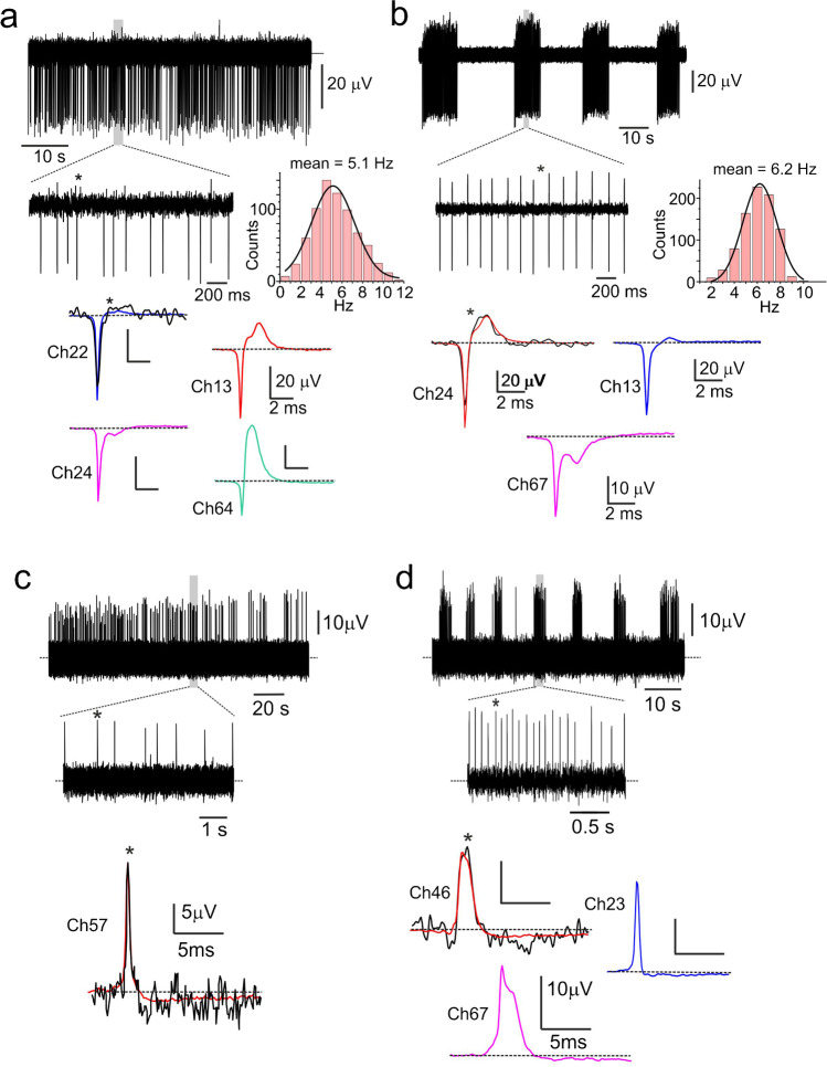
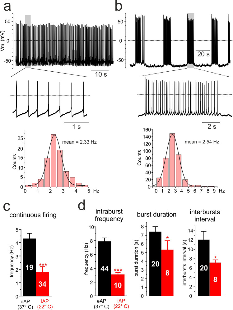
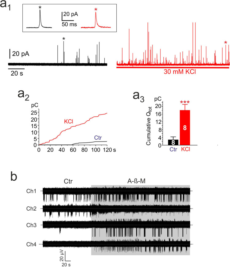
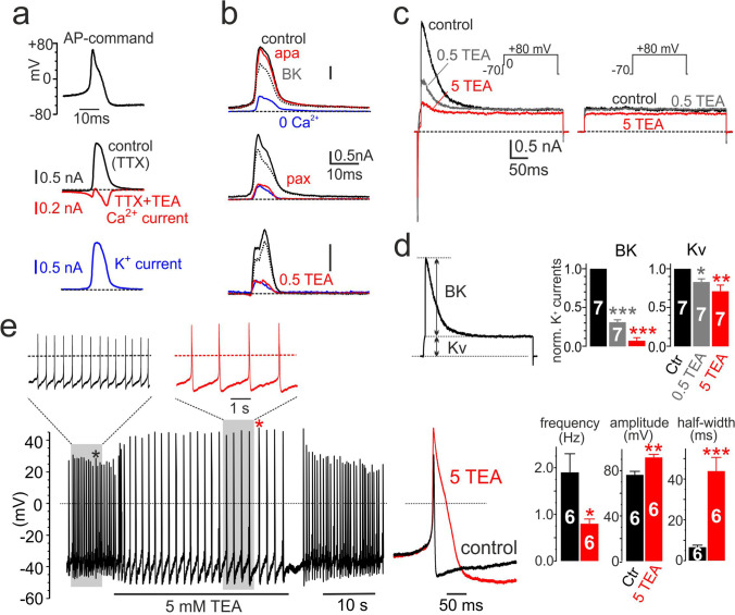
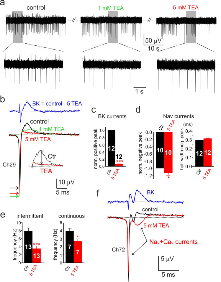
We recorded spontaneous extracellular action potentials (eAPs) from rat chromaffin cells (CCs) at 37 °C using microelectrode arrays (MEAs) and compared them with intracellularly recorded APs (iAPs) through conventional patch clamp recordings at 22 °C. We show the existence of two distinct firing modes on MEAs: a ~ 4 Hz irregular continuous firing and a frequent intermittent firing mode where periods of high-intraburst frequency (~ 8 Hz) of ~ 7 s duration are interrupted by silent periods of ~ 12 s. eAPs occurred either as negative- or positive-going signals depending on the contact between cell and microelectrode: either predominantly controlled by junction-membrane ion channels (negative-going) or capacitive/ohmic coupling (positive-going). Negative-going eAPs were found to represent the trajectory of the Na, Ca, and K currents passing through the cell area in tight contact with the microelectrode during an AP (point-contact junction). The inward Nav component of eAPs was blocked by TTX in a dose-dependent manner (IC ~ 10 nM) while the outward component was strongly attenuated by the BK channel blocker paxilline (200 nM) or TEA (5 mM). The SK channel blocker apamin (200 nM) had no effect on eAPs. Inward Nav and Cav currents were well-resolved after block of Kv and BK channels or in cells showing no evident outward K currents. Unexpectedly, on the same type of cells, we could also resolve inward L-type currents after adding nifedipine (3 μM). In conclusion, MEAs provide a direct way to record different firing modes of rat CCs and to estimate the Na, Ca, and K currents that sustain cell firing and spontaneous catecholamines secretion.
我们在 37°C 下使用微电极阵列(MEA)记录大鼠嗜铬细胞(CC)的自发细胞外动作电位(eAP),并通过在 22°C 下进行传统的膜片钳记录来比较细胞内记录的动作电位(iAP)。我们展示了 MEA 上存在两种不同的放电模式:一种约 4 Hz 的不规则连续放电和一种频繁的间歇性放电模式,其中高爆发频率(约 8 Hz)的持续时间约 7 s 的时期被约 12 s 的沉默期所打断。eAP 可以是负向或正向信号,这取决于细胞与微电极的接触:要么主要由连接膜离子通道(负向)控制,要么由电容/欧姆耦合(正向)控制。负向 eAP 被发现代表了在动作电位期间通过与微电极紧密接触的细胞区域的 Na、Ca 和 K 电流的轨迹(点接触连接)。eAP 的内向 Nav 成分被 TTX 以剂量依赖性方式阻断(IC 约 10 nM),而外向成分则被 BK 通道阻断剂 paxilline(200 nM)或 TEA(5 mM)强烈衰减。SK 通道阻断剂 apamin(200 nM)对 eAP 没有影响。在阻断 Kv 和 BK 通道或在没有明显外向 K 电流的细胞中,内向 Nav 和 Cav 电流得到很好的分辨。出乎意料的是,在同一类型的细胞中,我们还可以在添加硝苯地平(3 μM)后分辨内向 L 型电流。总之,MEA 提供了一种直接记录大鼠 CC 不同放电模式并估计维持细胞放电和自发性儿茶酚胺分泌的 Na、Ca 和 K 电流的方法。