Department of Ophthalmology, Columbia University, Vagelos College of Physicians and Surgeons, New York, NY, United States of America.
University of Michigan Medical School, Ann Arbor, MI, United States of America.
PLoS One. 2022 Oct 21;17(10):e0276629. doi: 10.1371/journal.pone.0276629. eCollection 2022.
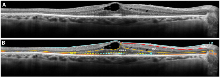
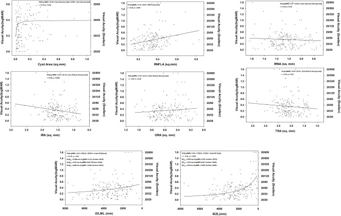
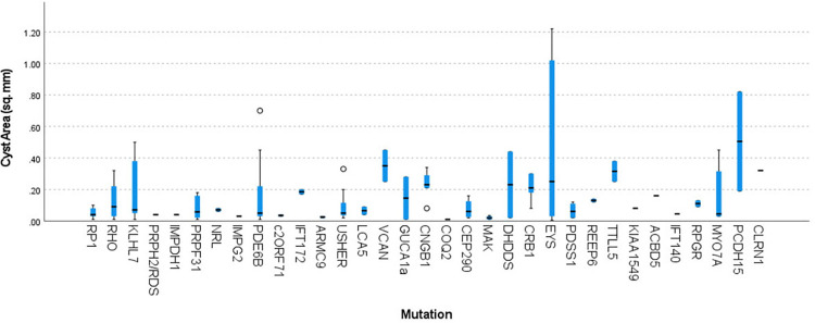
Cystoid macular edema (CME) is a major cause of central visual deterioration in retinitis pigmentosa. The exact reason for CME and its prognostic significance in this patient population is unknown. We seek to find clues to answer these questions by examining the anatomical correlations between retinal cysts and retinal morphometric parameters in a cohort of patients with retinitis pigmentosa and CME. For this reason, 103 patients (196 eyes) with untreated cystoid macular edema (CME) were identified from a pool of 578 genotyped patients with retinitis pigmentosa. Image analyses were conducted using three central horizontal OCT scans of these patients to calculate cross-sectional areas of the retinal nerve fiber layer, outer retinal, inner retinal, cysts, and total retinal areas. Lengths of the ellipsoid zone and outer limiting membrane were also measured. Best-fit curves were derived for analyzing the factors playing a role in the size of the retinal cysts and the patients' visual acuity. Generalized Estimating Equation and multivariate linear regression analyses were conducted to determine the correlations between visual acuity, morphometric and clinical data, and the significant cyst size and visual acuity determinants. Twenty-five percent of the screened patients (103/578) had CME. Patients with autosomal dominant retinitis pigmentosa had the highest incidence of CME (43.6%, p<0.001) but also had the best visual acuity (20/34±20/30, p = 0.02). The total cyst area was 0.14±0.18 mm2. Outer retinal area (B = 0.214; p = 0.008), age (B = -0.003; p<0.001) and retinal nerve fiber area (B = 0.411; p = 0.005) were main determinants of the (r = 0.44; p<0.001) cyst size. Cysts resolved with progressing retinal degeneration. Length of the intact ellipsoid zone (B = -5.16E-5; p<0.001), the inheritance pattern (B = 0.04; p = 0.028) and retinal nerve fiber area (B = 0.751; p<0.001) were the main determinants of visual acuity. In patients with retinitis pigmentosa and cystoid macular edema, retinal nerve fiber layer thickness is associated with decreasing visual acuity and cyst size. This finding suggests that intraretinal cysts may compress retinal axons and cause subsequent visual loss in retinitis pigmentosa.
囊样黄斑水肿(CME)是色素性视网膜炎中心视力恶化的主要原因。CME 的确切原因及其在该患者人群中的预后意义尚不清楚。我们通过检查一组色素性视网膜炎伴 CME 患者的视网膜囊肿与视网膜形态参数之间的解剖相关性,试图找到答案。为此,我们从 578 名经基因分型的色素性视网膜炎患者中确定了 103 名(196 只眼)未经治疗的囊样黄斑水肿(CME)患者。对这些患者的 3 个中央水平 OCT 扫描进行图像分析,以计算视网膜神经纤维层、外视网膜、内视网膜、囊肿和整个视网膜区域的横截面面积。还测量了椭圆体带和外节膜的长度。为分析影响视网膜囊肿大小和患者视力的因素,得出了最佳拟合曲线。采用广义估计方程和多元线性回归分析,确定了视力、形态计量学和临床数据之间的相关性,以及显著的囊肿大小和视力决定因素。在筛选的患者中,有 25%(103/578)患有 CME。常染色体显性遗传型色素性视网膜炎患者的 CME 发生率最高(43.6%,p<0.001),但视力也最好(20/34±20/30,p=0.02)。总的囊肿面积为 0.14±0.18mm2。外视网膜面积(B=0.214;p=0.008)、年龄(B=-0.003;p<0.001)和视网膜神经纤维面积(B=0.411;p=0.005)是囊肿大小的主要决定因素(r=0.44;p<0.001)。随着视网膜变性的进展,囊肿逐渐消退。完整的椭圆体带长度(B=-5.16E-5;p<0.001)、遗传模式(B=0.04;p=0.028)和视网膜神经纤维面积(B=0.751;p<0.001)是视力的主要决定因素。在色素性视网膜炎伴囊样黄斑水肿的患者中,视网膜神经纤维层厚度与视力下降和囊肿大小有关。这一发现表明,视网膜内囊肿可能压迫视网膜轴突,导致色素性视网膜炎的后续视力丧失。