QUEL Imaging, 85 N. Main Street Suite 142, White River Junction, VT, 05001, USA.
Thayer School of Engineering at Dartmouth, 14 Engineering Drive, Hanover, NH, 03755, USA.
Mol Imaging Biol. 2023 Feb;25(1):212-220. doi: 10.1007/s11307-022-01783-5. Epub 2022 Oct 28.
Interventional fluorescence imaging is increasingly being utilized to quantify cancer biomarkers in both clinical and preclinical models, yet absolute quantification is complicated by many factors. The use of optical phantoms has been suggested by multiple professional organizations for quantitative performance assessment of fluorescence guidance imaging systems. This concept can be further extended to provide standardized tools to compare and assess image analysis metrics.
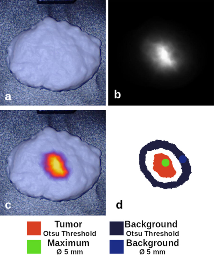
3D-printed fluorescence phantoms based on solid tumor models were developed with representative bio-mimicking optical properties. Phantoms were produced with discrete tumors embedded with an NIR fluorophore of fixed concentration and either zero or 3% non-specific fluorophore in the surrounding material. These phantoms were first imaged by two fluorescence imaging systems using two methods of image segmentation, and four assessment metrics were calculated to demonstrate variability in the quantitative assessment of system performance. The same analysis techniques were then applied to one tumor model with decreasing tumor fluorophore concentrations.
These anatomical phantom models demonstrate the ability to use 3D printing to manufacture anthropomorphic shapes with a wide range of reduced scattering (μ': 0.24-1.06 mm) and absorption (μ: 0.005-0.14 mm) properties. The phantom imaging and analysis highlight variability in the measured sensitivity metrics associated with tumor visualization.
3D printing techniques provide a platform for demonstrating complex biological models that introduce real-world complexities for quantifying fluorescence image data. Controlled iterative development of these phantom designs can be used as a tool to advance the field and provide context for consensus-building beyond performance assessment of fluorescence imaging platforms, and extend support for standardizing how quantitative metrics are extracted from imaging data and reported in literature.
介入荧光成像是一种越来越多地被用于量化临床和临床前模型中癌症生物标志物的方法,但由于许多因素的影响,绝对量化变得复杂。多个专业组织建议使用光学体模来评估荧光引导成像系统的定量性能。这一概念可以进一步扩展,为比较和评估图像分析指标提供标准化工具。
根据实体瘤模型开发了具有代表性的生物模拟光学特性的 3D 打印荧光体模。体模由离散肿瘤组成,内部嵌入固定浓度的近红外荧光染料,周围材料中要么没有非特异性荧光染料,要么含有 3%的非特异性荧光染料。使用两种荧光成像系统和两种图像分割方法对这些体模进行成像,计算了四个评估指标,以证明系统性能定量评估的可变性。然后,将相同的分析技术应用于一个肿瘤模型,该模型中的肿瘤荧光染料浓度逐渐降低。
这些解剖学体模模型证明了使用 3D 打印制造具有广泛降低散射(μ':0.24-1.06mm)和吸收(μ:0.005-0.14mm)特性的拟人形状的能力。体模成像和分析突出了与肿瘤可视化相关的测量灵敏度指标的可变性。
3D 打印技术为演示复杂的生物学模型提供了一个平台,这些模型引入了量化荧光图像数据的实际复杂性。这些体模设计的控制迭代开发可以作为一种工具,推动该领域的发展,并为超越荧光成像平台的性能评估建立共识提供背景,同时扩展对如何从成像数据中提取定量指标并在文献中报告的支持。