Dong Zhihong, Gong Jiabao, Zhang Haowei, Ni Yanting, Cheng Lijia, Song Qiaoyu, Tang Lu, Xing Fei, Liu Ming, Zhou Changchun
School of Mechanical Engineering, Chengdu University, Chengdu, 610106, China.
College of Electronics and Information Engineering, Sichuan University, Chengdu 610065, China.
Int J Bioprint. 2022 Sep 1;8(4):613. doi: 10.18063/ijb.v8i4.613. eCollection 2022.
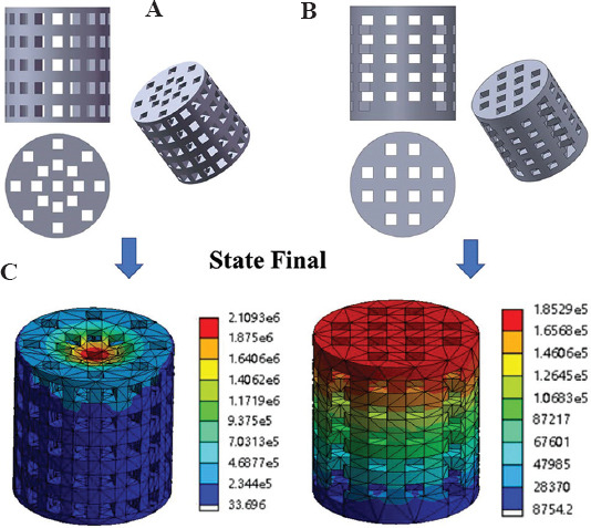
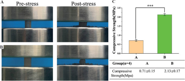
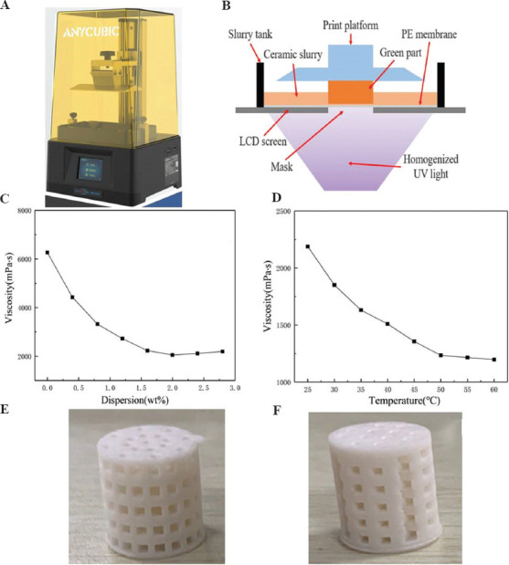
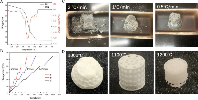
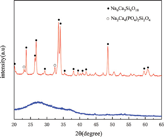
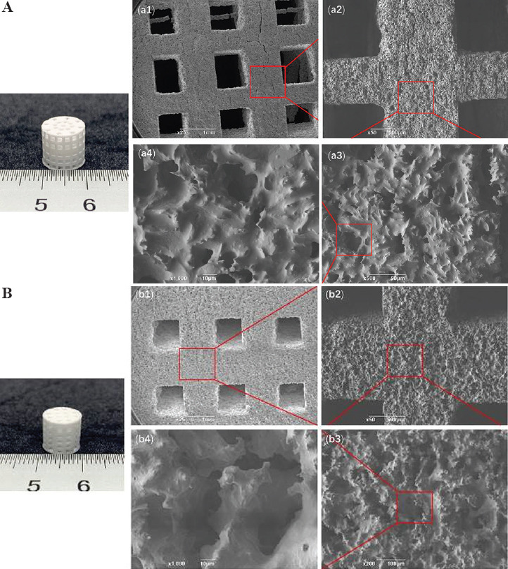
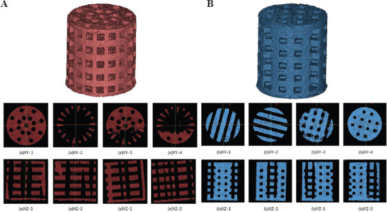
Three-dimensional (3D) printing technology provides advanced technical support for designing personalized bone tissue engineering scaffold. In this study, two porous diffusing models, namely, average and layered perforated cylindrical scaffolds, were designed for bone tissue engineering scaffold. The designed models were fabricated by liquid crystal display mask stereolithography printing. Structural design and finite element mechanical analysis were conducted. 45S5 bioglass was selected as the raw material for preparing the printing inks for bone tissue engineering scaffolds. By adjusting the viscosity and temperature of the slurry, the maximum proportion of 45S5 bioglass (40 wt%) was added into the photosensitive resin for preparing 3D printing slurry. Our results indicated that an optimized sintering condition includes the debinding rate (0.5°C/min), and temperature raising rate (5°C/min) and sintering temperature (1100°C) were proposed to sinter 45S5 bioceramic scaffolds. The amorphous 45S5 bioglass showed good crystallization after sintering, and the scaffold porous structure showed good integrity. Micropores were observed in the struts which interconnected with each other. Moreover, the porosities were tested as 57% and 45% with a uniform pore distribution. The shrinkage rate was about 10% during sintering process due to binder burning and crystallization shrinkage. The compressive strength of the sintered scaffold was 0.71 ± 0.048 MPa and 2.13 ± 0.054 MPa, respectively, which are consistent with the finite element mechanical analysis simulation results. In conclusion, the layered perforated 45S5 bioglass scaffold shows good mechanical properties and porosity, indicating that it could be a promising candidate for bone tissue engineering.
三维(3D)打印技术为设计个性化骨组织工程支架提供了先进的技术支持。在本研究中,为骨组织工程支架设计了两种多孔扩散模型,即平均多孔和分层多孔圆柱形支架。所设计的模型通过液晶显示掩膜立体光刻印刷制造。进行了结构设计和有限元力学分析。选择45S5生物玻璃作为制备骨组织工程支架打印油墨的原材料。通过调节浆料的粘度和温度,将45S5生物玻璃的最大比例(40 wt%)添加到光敏树脂中以制备3D打印浆料。我们的结果表明,提出了优化的烧结条件,包括脱脂速率(0.5°C/分钟)、升温速率(5°C/分钟)和烧结温度(1100°C)来烧结45S5生物陶瓷支架。非晶态的45S5生物玻璃在烧结后显示出良好的结晶,并且支架多孔结构显示出良好的完整性。在相互连接的支柱中观察到微孔。此外,测试得到孔隙率分别为57%和45%,且孔隙分布均匀。由于粘结剂燃烧和结晶收缩,烧结过程中的收缩率约为10%。烧结支架的抗压强度分别为0.71±0.048 MPa和2.13±0.054 MPa,这与有限元力学分析模拟结果一致。总之,分层多孔45S5生物玻璃支架显示出良好的力学性能和孔隙率,表明它可能是骨组织工程的一个有前途的候选材料。