Centre for Clinical Brain Sciences, University of Edinburgh, Edinburgh, UK.
Anne Rowling Regenerative Neurology Clinic, University of Edinburgh, Edinburgh, UK.
Sci Rep. 2022 Nov 28;12(1):20472. doi: 10.1038/s41598-022-24312-4.
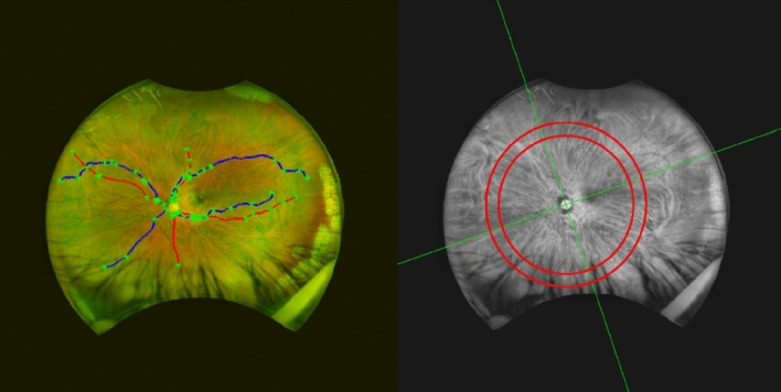
Our purpose was to investigate changes to the retina in multiple sclerosis (MS) using established and novel modes of retinal image acquisition and analysis. 72 participants with MS and 80 healthy volunteers underwent retinal scanning with optical coherence tomography (OCT) and ultra-widefield (UWF) scanning laser ophthalmoscopy (SLO), over a two-year period. Changes in retinal nerve fibre layer (RNFL) thickness, macular volume and retinal blood vessel diameter were measured and parameters were then tested for associations with MS. Measurements from OCT showed that individuals with MS had a thinner RNFL and reduced macular volume when compared to healthy volunteers. On UWF images, participants with MS had reduced arterial widths in the inferior nasal quadrant of both eyes and reduced venous widths in the inferior nasal quadrant of right eyes. Longitudinal analysis showed that participants with MS had an accelerated annual rate of RNFL thinning in several regions of the retina. In conclusion, the assessment of OCT showed thinning of the RNFL and macula in concordance with previous reports on MS, while analysis of blood vessels in the retinal periphery from UWF-SLO images revealed novel changes.
我们的目的是使用已建立的和新的视网膜图像采集和分析模式来研究多发性硬化症(MS)中的视网膜变化。在两年的时间里,72 名 MS 患者和 80 名健康志愿者接受了光学相干断层扫描(OCT)和超广角(UWF)扫描激光检眼镜(SLO)的视网膜扫描。测量了视网膜神经纤维层(RNFL)厚度、黄斑体积和视网膜血管直径的变化,并测试了这些参数与 MS 的相关性。OCT 的测量结果表明,与健康志愿者相比,MS 患者的 RNFL 更薄,黄斑体积更小。在 UWF 图像上,MS 患者的双眼下鼻象限动脉变窄,右眼下鼻象限静脉变窄。纵向分析显示,MS 患者的几个视网膜区域的 RNFL 变薄速度加快。总之,OCT 的评估结果显示,RNFL 和黄斑变薄与之前关于 MS 的报告一致,而 UWF-SLO 图像中对视网膜周边血管的分析则揭示了新的变化。