Clinical Neurophysiology Research Laboratory, Department of Psychiatry, Western Psychiatric Hospital, University of Pittsburgh School of Medicine, Pittsburgh, PA, USA.
Clinical Neurophysiology Research Laboratory, Department of Psychiatry, Western Psychiatric Hospital, University of Pittsburgh School of Medicine, Pittsburgh, PA, USA.
Neuroimage Clin. 2022;36:103261. doi: 10.1016/j.nicl.2022.103261. Epub 2022 Nov 7.
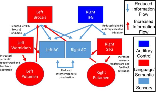
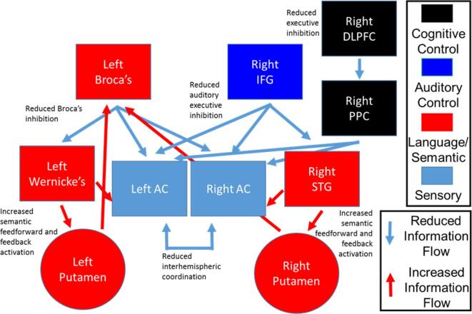
Cortical (e.g., Broca's area and Wernicke's area) and subcortical (e.g., putamen) language-related areas and executive control areas (e.g., inferior frontal gyrus (IFG), dorsolateral prefrontal cortex (DLPFC)) show functional and structural dysconnectivity in long-term psychosis. We examined whether resting-state basal perfusion levels revealed selective pathophysiology (likely hypo- and hyper-activation) of language-related and executive areas in first-episode psychosis (FEP).
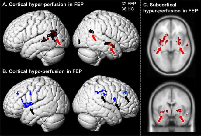
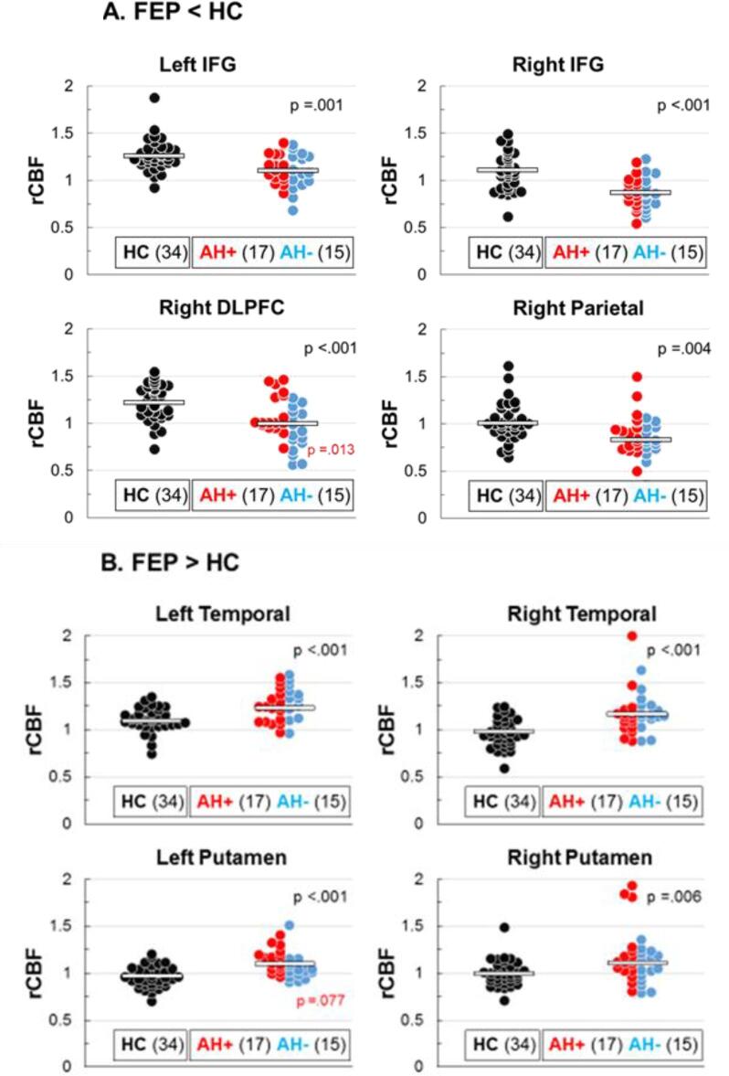
Basal resting-state perfusion was measured using pseudo-continuous Arterial Spin Labeling (pcASL). Relative cerebral blood flow (rCBF) was compared between 32 FEP and 34 matched healthy comparison (HC) individuals. Structural and functional MRI scans were acquired using a 3T Prisma scanner during the same session.
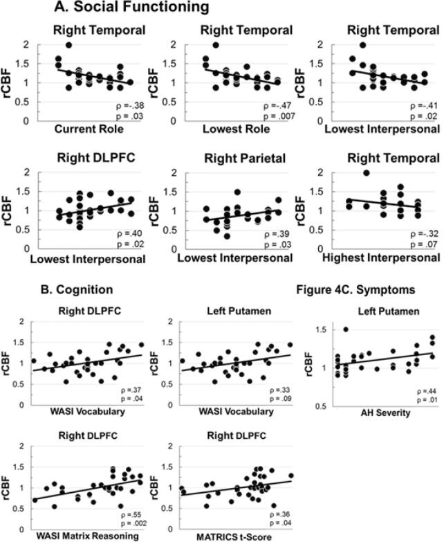
Whole-brain comparison of resting rCBF identified 8 clusters with significant between-group differences. Reduced rCBF was found in executive control areas in left and right IFG, right DLPFC, and right parietal cortex. Increased rCBF was found in left and right temporal cortex (including Wernicke's area), and left and right putamen. A positive correlation was observed between auditory hallucination severity and rCBF in the left putamen.
To the degree that perfusion implies activation, language and auditory processing areas in bilateral temporal lobe and putamen showed pathological hyper-activity, and cognitive control areas (IFG, DLPFC, right parietal) showed pathological hypo-activity in FEP at rest. Pathological basal activity was present across the range of symptom severity, suggesting it may be a common underlying pathology for psychosis that may be targeted with non-invasive brain stimulation to normalize resting activity levels.
皮质(例如,布罗卡区和韦尼克区)和皮质下(例如,壳核)语言相关区域和执行控制区域(例如,额下回(IFG),背外侧前额叶皮层(DLPFC))在长期精神病中显示功能和结构连接不良。我们研究了初发性精神病(FEP)患者静息状态下的基础灌注水平是否揭示了语言相关和执行区域的选择性病理生理学(可能是低激活和高激活)。
使用伪连续动脉自旋标记(pcASL)测量基础静息状态灌注。将 32 名 FEP 患者和 34 名匹配的健康对照(HC)个体的相对脑血流(rCBF)进行比较。在同一时间段内,使用 3T Prisma 扫描仪采集结构和功能 MRI 扫描。
全脑比较静息 rCBF 发现 8 个组间差异有统计学意义的簇。在左、右 IFG、右 DLPFC 和右顶叶皮质的执行控制区域中发现 rCBF 减少。在左、右颞叶(包括韦尼克区)和左、右壳核中发现 rCBF 增加。在左壳核中,听觉幻觉严重程度与 rCBF 之间存在正相关。
在某种程度上,灌注暗示激活,双侧颞叶和壳核的语言和听觉处理区域表现出病理性高活动,而认知控制区域(IFG、DLPFC、右顶叶)在 FEP 静息时表现出病理性低活动。在症状严重程度的范围内存在病理性基础活动,这表明它可能是精神病的一种常见潜在病理,可能可以通过非侵入性脑刺激来靶向,以正常化静息活动水平。