Önnheim Karin, Huang Shan, Strid Holmertz Alexander, Andersson Sofia, Lönnblom Erik, Jonsson Charlotte, Holmdahl Rikard, Gjertsson Inger
Dep of Rheumatology and Inflammation Research, Institute of Medicine, Gothenburg University, Sweden.
Medical Inflammation Research, Dept of Medical Biochemistry and Biophysics, the Karolinska Institute, Stockholm, Sweden.
Osteoarthr Cartil Open. 2022 Jan 21;4(1):100235. doi: 10.1016/j.ocarto.2022.100235. eCollection 2022 Mar.
To investigate whether articular chondrocytes from rheumatoid arthritis (RA) patients have acquired a proinflammatory phenotype.
Articular cartilage explants from RA patients and healthy controls (HC) were cultured with or without interleukin (IL)-1β for two weeks. Protein levels of cytokines and metalloproteinases (MMPs) in the supernatant were measured by LUMINEX, mRNA with qPCR and nitrogen oxide (NO) levels with Griess assay.
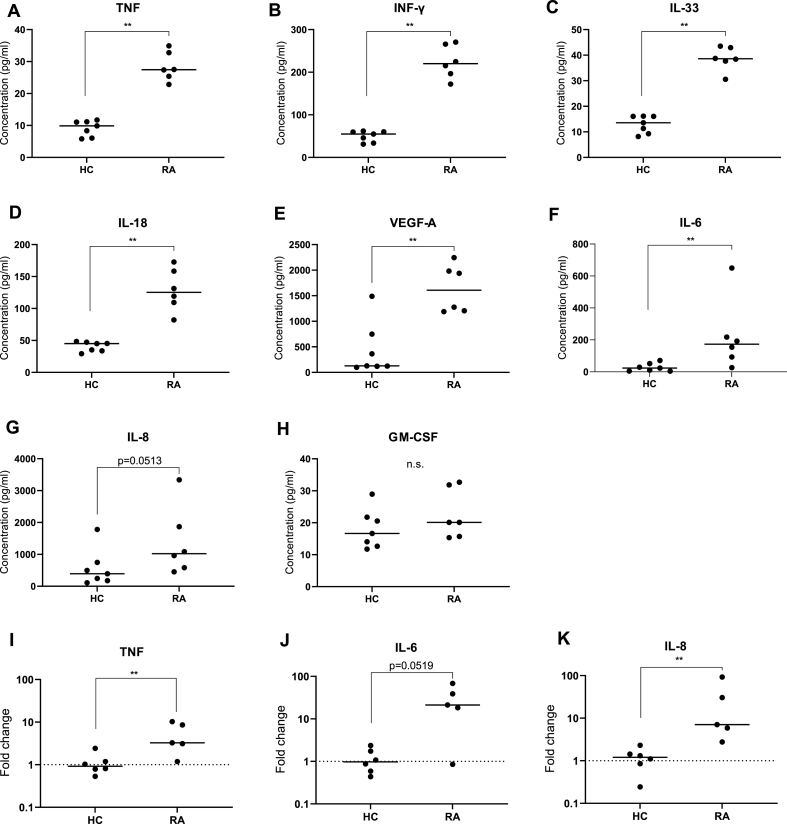
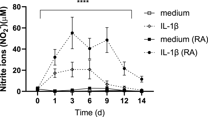
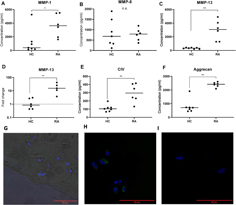
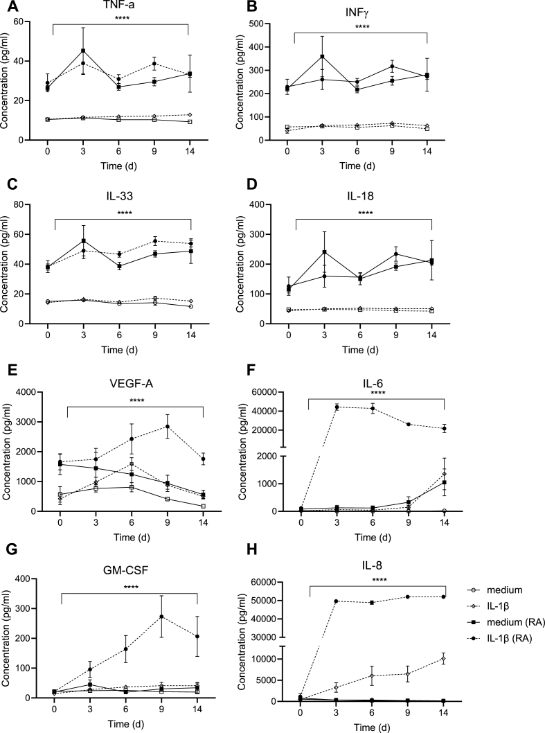
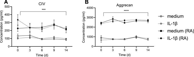
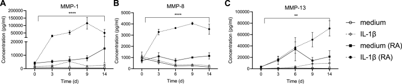
Within 24 h after culture, cartilage explants from RA spontaneously produced MMP-1 and MMP-13, and matrix components (aggrecan and collagen type IV) were released. In addition, the RA explants released higher levels of tumor necrosis factor, interferon-γ, IL-33, IL-18, vascular endothelial growth factor-A, IL-6 but not IL-8, and granulocyte-macrophage colony-stimulating factor (GM-CSF) as compared with HC. During two weeks of incubation the higher levels did not diminish. IL-1β stimulation further increased the levels of IL-6, IL-8 and GM-CSF, mainly in RA explants, and induced increased levels of NO in the supernatant from both HC and RA explants, as a result of chondrocyte activation.
RA chondrocytes are activated with a proinflammatory profile involving the production of cytokines as well as MMP-1 and MMP-13, that can lead to release of matrix molecules after activation, which suggests that the chondrocytes have a proinflammatory phenotype and thereby an active role in the pathogenesis.
研究类风湿关节炎(RA)患者的关节软骨细胞是否获得了促炎表型。
将RA患者和健康对照者(HC)的关节软骨外植体在有或无白细胞介素(IL)-1β的情况下培养两周。通过LUMINEX检测上清液中细胞因子和金属蛋白酶(MMPs)的蛋白水平,用qPCR检测mRNA水平,用Griess法检测一氧化氮(NO)水平。
培养后24小时内,RA患者的软骨外植体自发产生MMP-1和MMP-13,并释放基质成分(聚集蛋白聚糖和IV型胶原)。此外,与HC相比,RA外植体释放的肿瘤坏死因子、干扰素-γ、IL-33、IL-18、血管内皮生长因子-A、IL-6水平更高,但IL-8和粒细胞-巨噬细胞集落刺激因子(GM-CSF)水平不高。在两周的孵育过程中,较高水平并未降低。IL-1β刺激进一步增加了IL-6、IL-8和GM-CSF的水平,主要是在RA外植体中,并导致HC和RA外植体上清液中的NO水平升高,这是软骨细胞活化的结果。
RA软骨细胞被激活,具有涉及细胞因子以及MMP-1和MMP-13产生的促炎特征,激活后可导致基质分子释放,这表明软骨细胞具有促炎表型,从而在发病机制中起积极作用。