Hernández-Jiménez Tania, Cruz-Nova Pedro, Ancira-Cortez Alejandra, Gibbens-Bandala Brenda, Lara-Almazán Nancy, Ocampo-García Blanca, Santos-Cuevas Clara, Morales-Avila Enrique, Ferro-Flores Guillermina
Department of Radioactive Materials, Instituto Nacional de Investigaciones Nucleares, Ocoyoacac 52750, Mexico.
Faculty of Chemistry, Universidad Autónoma del Estado de México, Toluca 50180, Mexico.
Nanomaterials (Basel). 2022 Nov 25;12(23):4181. doi: 10.3390/nano12234181.
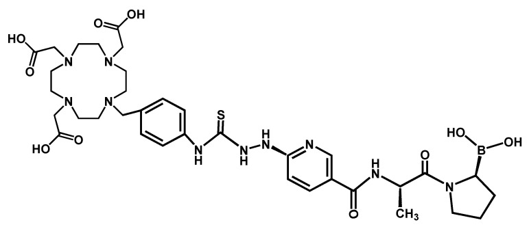
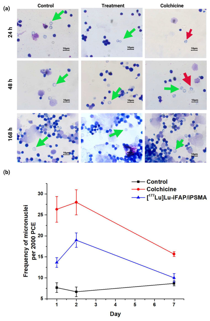
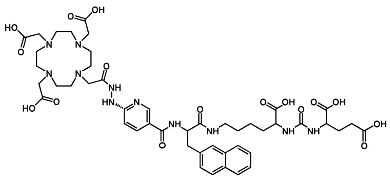
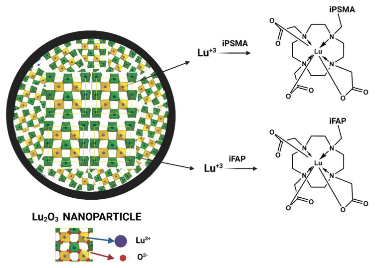
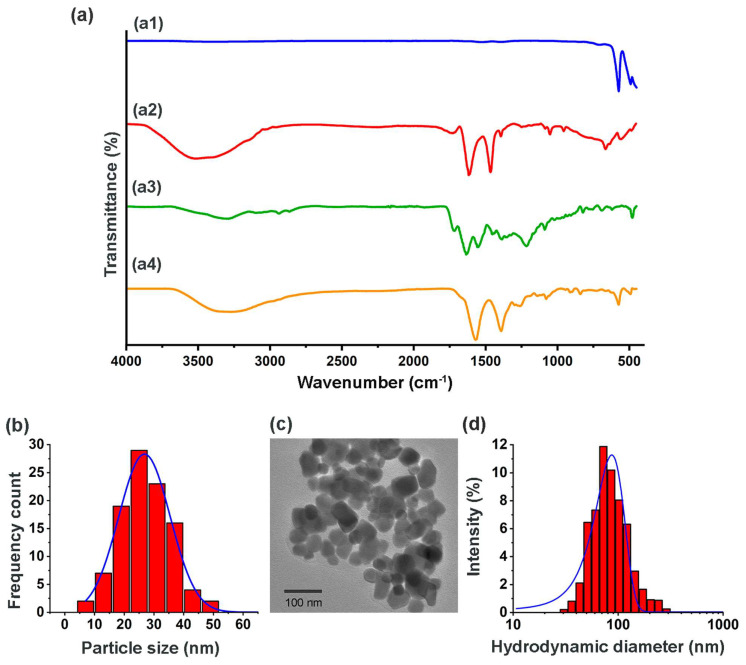
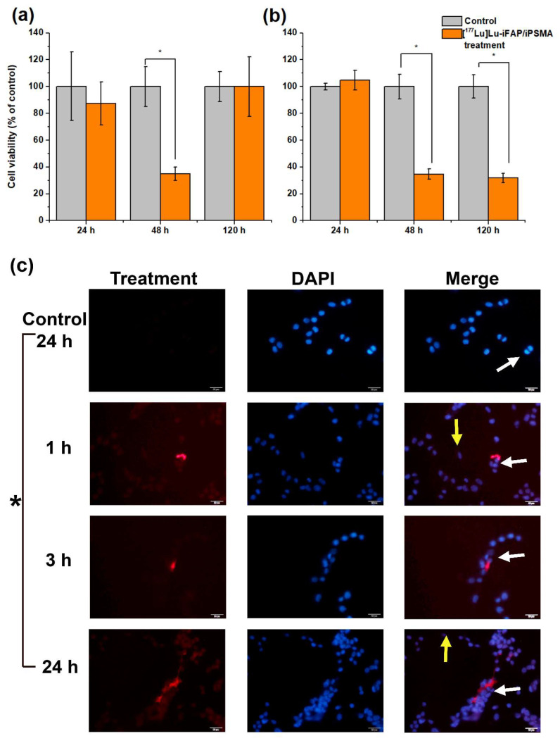
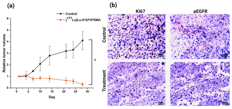
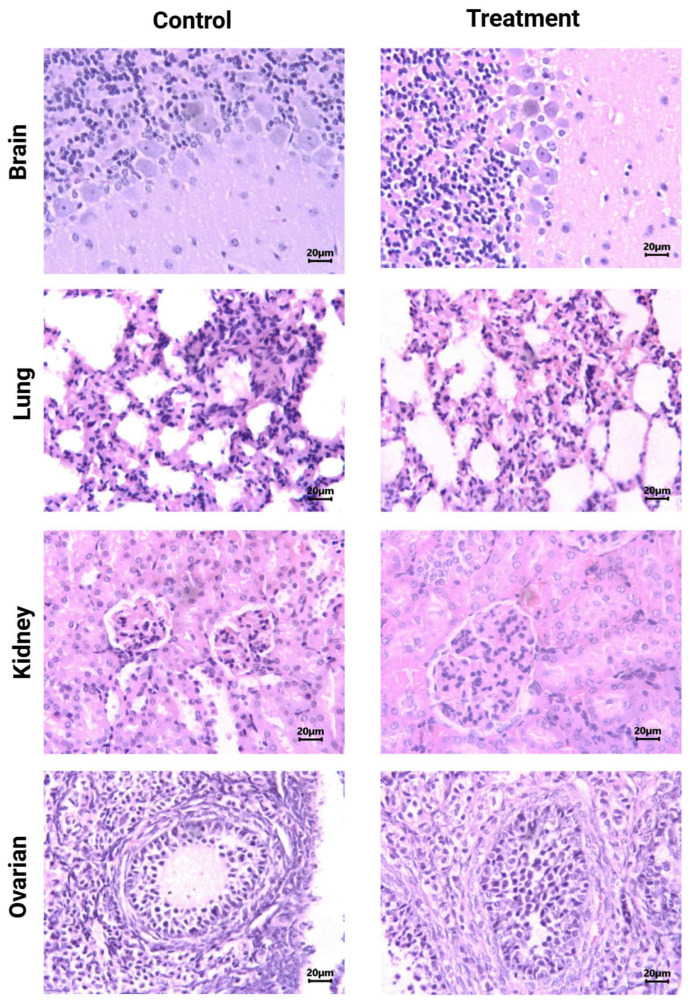
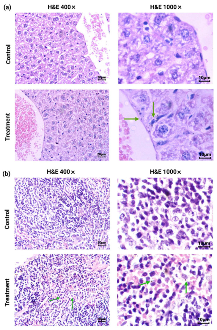
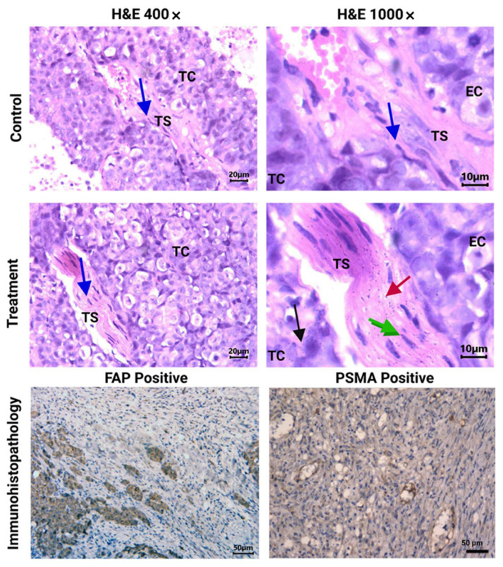
The fibroblast activation protein (FAP) is heavily expressed in fibroblasts associated with the tumor microenvironment, while the prostate-specific membrane antigen (PSMA) is expressed in the neovasculature of malignant angiogenic processes. Previously, we reported that [Lu]lutetium sesquioxide-iFAP/iPSMA nanoparticles ([Lu]Lu-iFAP/iPSMA) inhibit HCT116 tumor progression in mice. Understanding the toxicity of [Lu]Lu-iFAP/iPSMA in healthy tissues, as well as at the tissue and cellular level in pathological settings, is essential to demonstrate the nanosystem safety for treating patients. It is equally important to demonstrate that [Lu]Lu-iFAP/iPSMA can be prepared under good manufacturing practices (GMP) with reproducible pharmaceutical-grade quality characteristics. This research aimed to prepare [Lu]Lu-iFAP/iPSMA under GMP-compliant radiopharmaceutical processes and evaluate its toxicity in cell cultures and murine biological systems under pathological environments. [Lu]LuO nanoparticles were formulated as radiocolloidal solutions with FAP and PSMA inhibitor ligands (iFAP and iPSMA), sodium citrate, and gelatin, followed by heating at 121 °C (103-kPa pressure) for 15 min. Three consecutive batches were manufactured. The final product was analyzed according to conventional pharmacopeial methods. The Lu content in the formulations was determined by X-ray fluorescence. [Lu]Lu-iFAP/iPSMA performance in cancer cells was evaluated in vitro by immunofluorescence. Histopathological toxicity in healthy and tumor tissues was assessed in HCT116 tumor-bearing mice. Immunohistochemical assays were performed to corroborate FAP and PSMA tumor expression. Acute genotoxicity was evaluated using the micronuclei assay. The results showed that the batches manufactured under GMP conditions were reproducible. Radiocolloidal solutions were sterile and free of bacterial endotoxins, with radionuclidic and radiochemical purity greater than 99%. The lutetium content was 0.10 ± 0.02 mg/mL (0.9 GBq/mg). Significant inhibition of cell proliferation in vitro and in tumors was observed due to the accumulation of nanoparticles in the fibroblasts (FAP+) and neovasculature (PSMA+) of the tumor microenvironment. No histopathological damage was detected in healthy tissues. The data obtained in this research provide new evidence on the selective toxicity to malignant tumors and the absence of histological changes in healthy tissues after intravenous injection of [Lu]Lu-iFAP/iPSMA in mammalian hosts. The easy preparation under GMP conditions and the toxicity features provide the added value needed for [Lu]Lu-iFAP/iPSMA clinical translation.
成纤维细胞激活蛋白(FAP)在与肿瘤微环境相关的成纤维细胞中大量表达,而前列腺特异性膜抗原(PSMA)则在恶性血管生成过程的新生血管中表达。此前,我们报道了[镥]氧化镥-iFAP/iPSMA纳米颗粒([镥]Lu-iFAP/iPSMA)可抑制小鼠体内HCT116肿瘤的进展。了解[镥]Lu-iFAP/iPSMA在健康组织以及病理环境下的组织和细胞水平的毒性,对于证明该纳米系统治疗患者的安全性至关重要。同样重要的是要证明[镥]Lu-iFAP/iPSMA可以按照良好生产规范(GMP)制备,具有可重现的药用级质量特性。本研究旨在按照符合GMP的放射性药物制备工艺制备[镥]Lu-iFAP/iPSMA,并评估其在病理环境下细胞培养和小鼠生物系统中的毒性。将[镥]LuO纳米颗粒与FAP和PSMA抑制剂配体(iFAP和iPSMA)、柠檬酸钠和明胶配制成放射性胶体溶液,然后在121°C(103 kPa压力)下加热15分钟。连续制备了三批产品。最终产品按照传统药典方法进行分析。通过X射线荧光法测定制剂中的镥含量。通过免疫荧光法在体外评估[镥]Lu-iFAP/iPSMA在癌细胞中的性能。在荷HCT116肿瘤的小鼠中评估健康组织和肿瘤组织中的组织病理学毒性。进行免疫组织化学分析以证实FAP和PSMA在肿瘤中的表达。使用微核试验评估急性遗传毒性。结果表明,在GMP条件下制备的批次具有可重复性。放射性胶体溶液无菌且无细菌内毒素,放射性核素和放射化学纯度均大于99%。镥含量为0.10±0.02 mg/mL(0.9 GBq/mg)。由于纳米颗粒在肿瘤微环境的成纤维细胞(FAP+)和新生血管(PSMA+)中积累,在体外和肿瘤中均观察到细胞增殖受到显著抑制。在健康组织中未检测到组织病理学损伤。本研究获得的数据为静脉注射[镥]Lu-iFAP/iPSMA后对哺乳动物宿主恶性肿瘤的选择性毒性以及健康组织中无组织学变化提供了新的证据。在GMP条件下易于制备以及毒性特征为[镥]Lu-iFAP/iPSMA的临床转化提供了所需的附加值。