Department of Microbiology and Immunology, Columbia University Irving Medical Center, New York, NY, USA.
J Exp Med. 2023 Mar 6;220(3). doi: 10.1084/jem.20221462. Epub 2022 Dec 19.
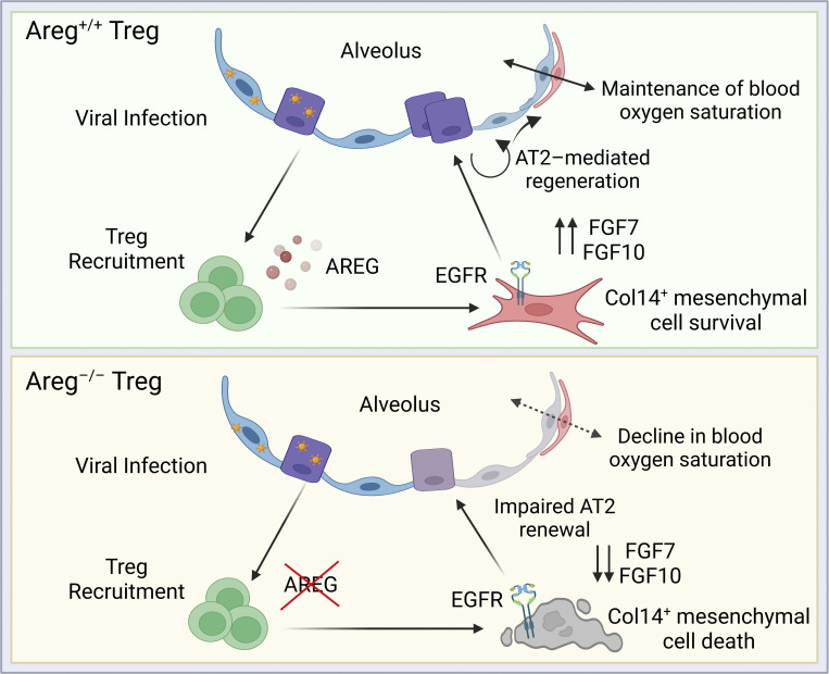
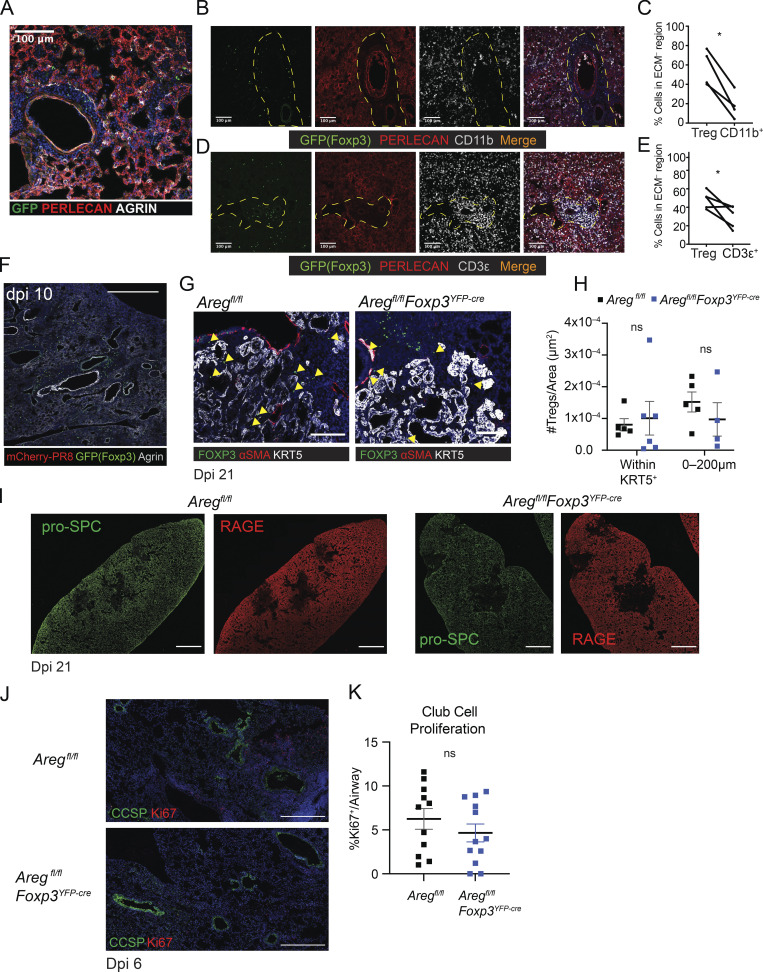
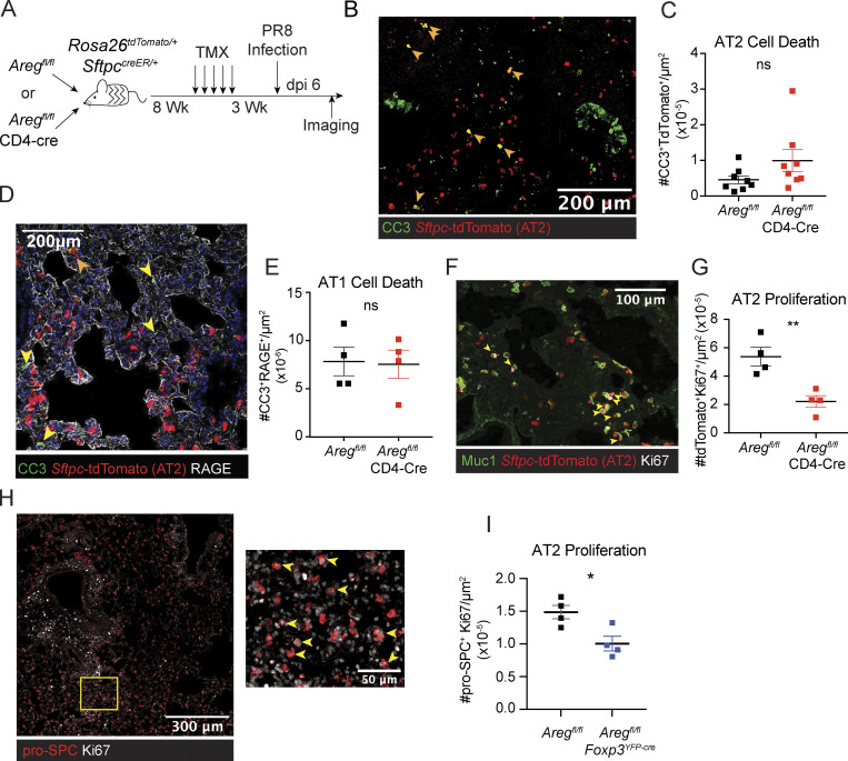
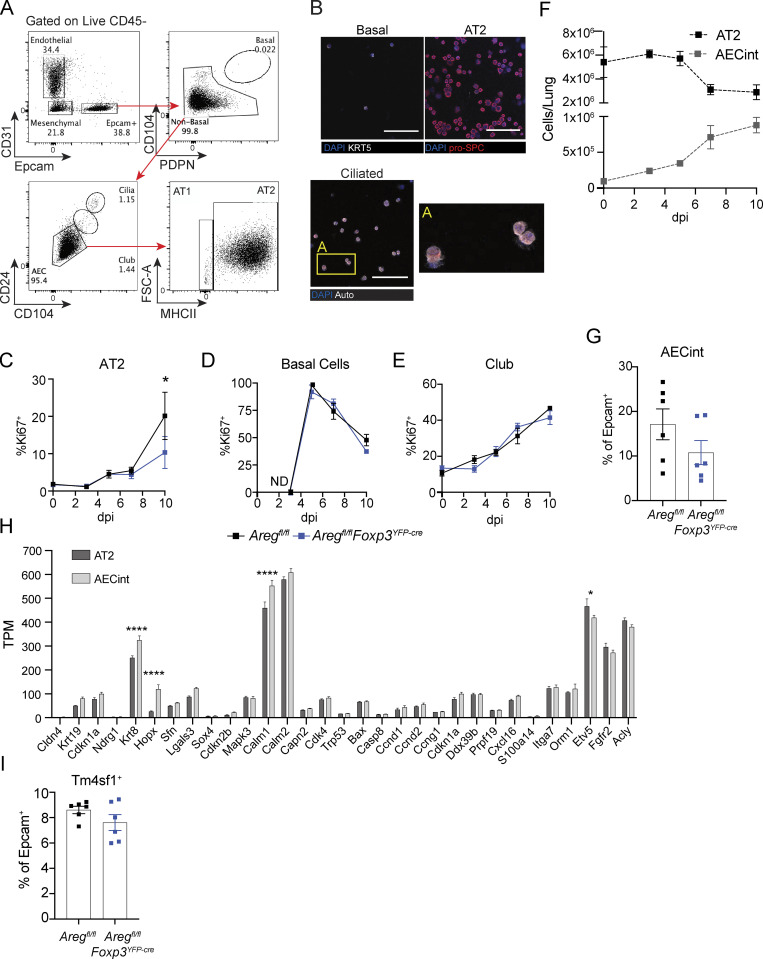
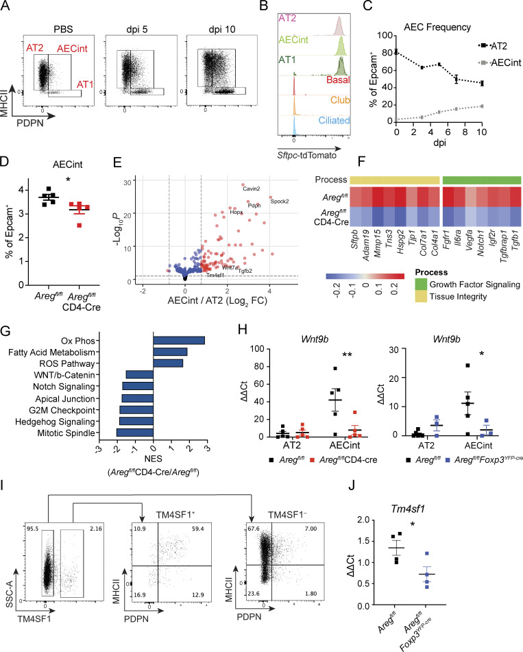
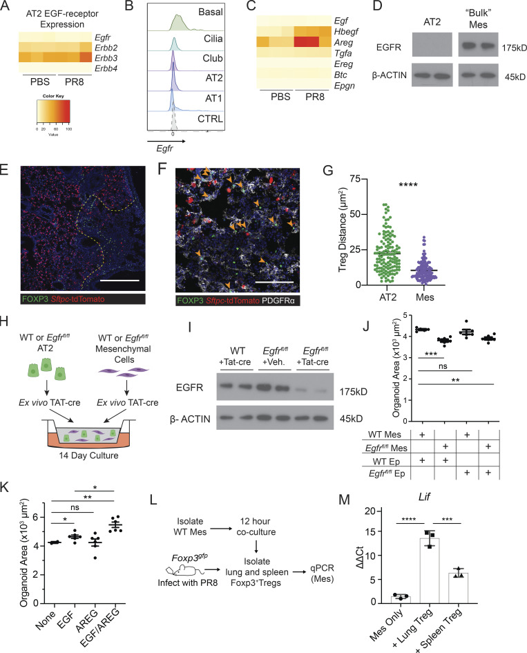
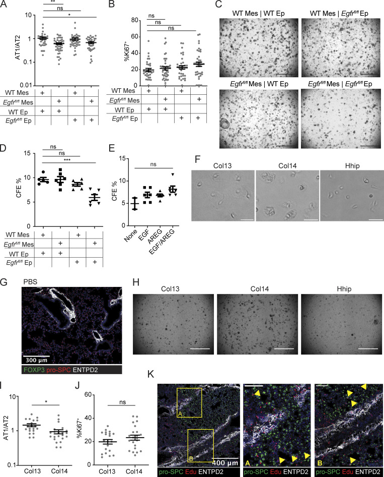
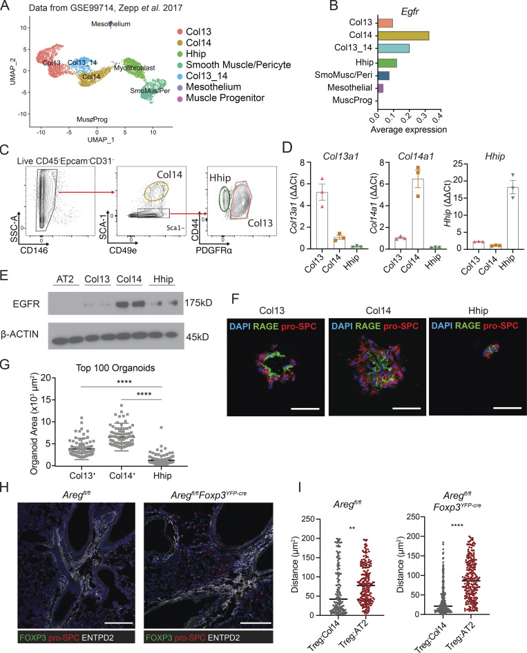
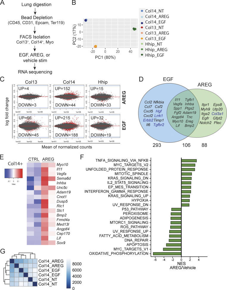
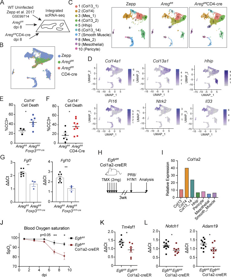
Following respiratory viral infection, regeneration of the epithelial barrier is required to preserve lung function and prevent secondary infections. Lung regulatory T (Treg) cells are critical for maintaining blood oxygenation following influenza virus infection through production of the EGFR ligand amphiregulin (Areg); however, how Treg cells engage with progenitors within the alveolar niche is unknown. Here, we describe local interactions between Treg cells and an Areg-responsive population of Col14a1+EGFR+ lung mesenchymal cells that mediate type II alveolar epithelial (AT2) cell-mediated regeneration following influenza virus infection. We propose a mechanism whereby Treg cells are deployed to sites of damage and provide pro-survival cues that support mesenchymal programming of the alveolar niche. In the absence of fibroblast EGFR signaling, we observe impaired AT2 proliferation and disrupted lung remodeling following viral clearance, uncovering a crucial immune/mesenchymal/epithelial network that guides alveolar regeneration.
呼吸道病毒感染后,需要再生上皮屏障以维持肺功能和防止继发感染。在流感病毒感染后,肺调节性 T (Treg) 细胞通过产生表皮生长因子受体 (EGFR) 配体 Amphiregulin (Areg) 对于维持血氧合至关重要;然而,Treg 细胞如何与肺泡龛内的祖细胞相互作用尚不清楚。在这里,我们描述了 Treg 细胞与 Areg 反应性 Col14a1+EGFR+肺间充质细胞之间的局部相互作用,这些细胞介导流感病毒感染后 II 型肺泡上皮 (AT2) 细胞介导的再生。我们提出了一种机制,即 Treg 细胞被部署到损伤部位,并提供支持肺泡龛间质编程的生存信号。在没有成纤维细胞 EGFR 信号的情况下,我们观察到病毒清除后 AT2 增殖受损和肺重塑中断,揭示了指导肺泡再生的重要免疫/间充质/上皮网络。