Department of Respiratory and Critical Care Medicine, Zhongshan Hospital, Fudan University, Shanghai Institute of Respiratory Disease, Shanghai, 200032, China.
Department of Respiratory and Critical Care Medicine, Minhang Hospital, Fudan University, Shanghai, 201199, China.
Respir Res. 2022 Dec 22;23(1):372. doi: 10.1186/s12931-022-02276-3.
To investigate the prognostic value of quantitative analysis of CT among patients with idiopathic pulmonary fibrosis (IPF) by quantifying the fibrosis extent and to attempt to provide precise medium-long term prognostic predictions for individual patients.
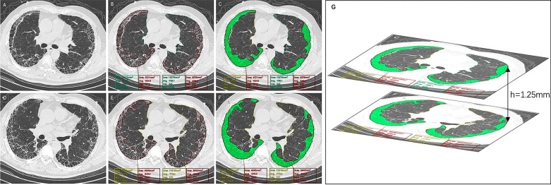
This was a retrospective cohort study that included 95 IPF patients in Zhongshan Hospital, Fudan University. 64 patients firstly diagnosed with IPF from 2009 to 2015 was included as the derivation cohort. Information regarding sex, age, the Gender-Age-Physiology (GAP) index, high-resolution computed tomography (HRCT) images, survival status, and pulmonary function parameters including forced vital capacity (FVC), FVC percent predicted (FVC%pred), diffusing capacity of carbon monoxide (DLCO), DLCO percent predicted (DLCO%pred), carbon monoxide transfer coefficient (KCO), KCO percent predicted (KCO%pred) were collected. 31 patients were included in the validation cohort. The Synapse 3D software was used to quantify the fibrotic lung volume (FLV) and total lung volume (TLV). The ratio of FLV to TLV was calculated and labeled CT, reflecting the extent of fibrosis. All the physiological variants and CT were analyzed for the dimension of survival through both univariate analysis and multivariate analysis. Formulas for predicting the probability of death based on the baseline CT were calculated by logistic regression, and validated by the validation cohort.
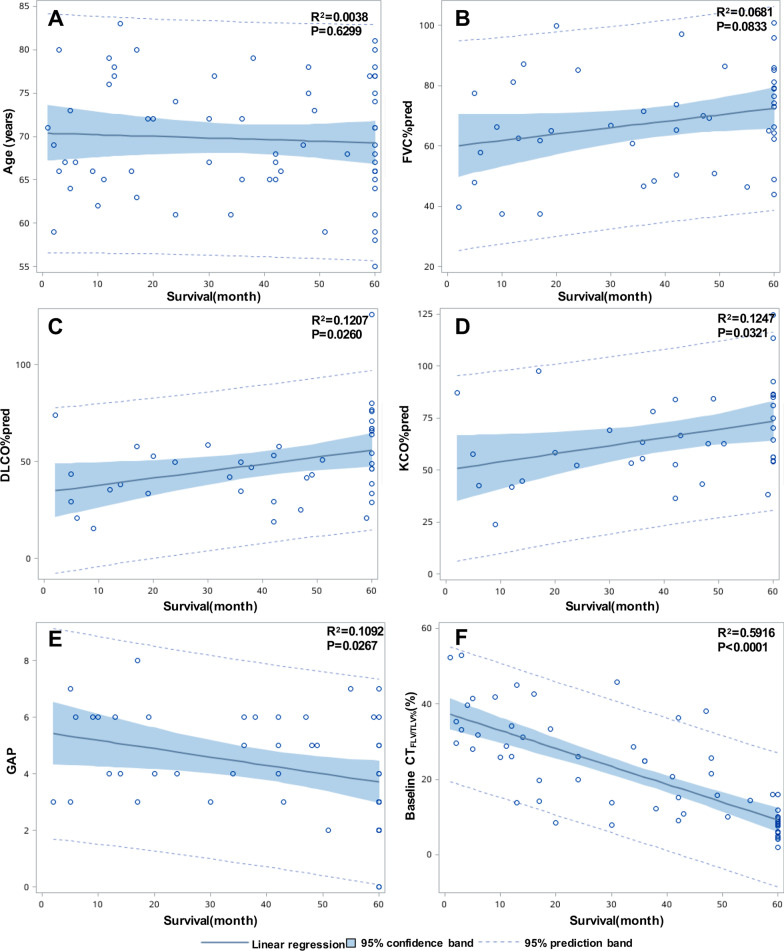
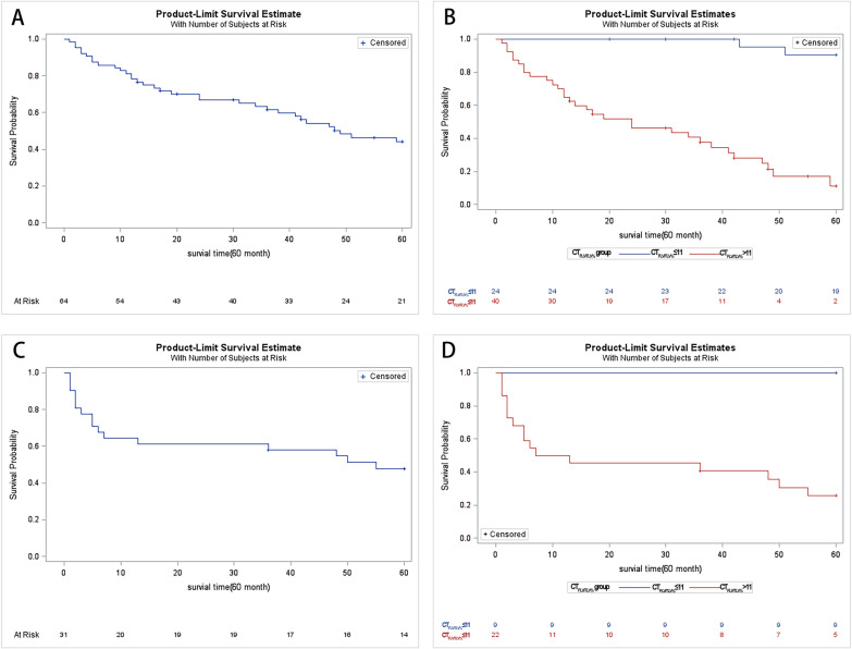
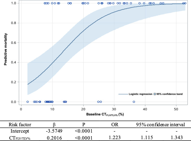
The univariate analysis indicated that CT along with DLCO%pred, KCO%pred and GAP index were significantly correlated with survival. However, only CT was meaningful in the multivariate analysis for prognostic prediction (HR 1.114, 95% CI 1.047-1.184, P = 0.0006), and the best cutoff was 11%, based on receiver operating characteristic (ROC) curve analysis. The survival times for the CT ≤ 11% and CT > 11% groups were significantly different. Given the CT data, the death probability of a patient at 1 year, 3 years and 5 years could be calculated by using a particular formula. The formulas were tested by the validation cohort, showed high sensitivity (88.2%), specificity (92.8%) and accuracy (90.3%).
Quantitative volume analysis of CT might be useful for evaluating the extent of fibrosis in the lung. The CT could be a valuable biomarker for precisely predicting the medium-long term prognosis of individual patients with IPF.
通过定量分析特发性肺纤维化(IPF)患者的 CT 来探讨 CT 对 IPF 患者的预后价值,试图为个体患者提供更准确的中远期预后预测。
这是一项回顾性队列研究,纳入了复旦大学中山医院的 95 名 IPF 患者。将 2009 年至 2015 年首次诊断为 IPF 的 64 名患者纳入为推导队列。收集了患者的性别、年龄、性别-年龄-生理评分(GAP)指数、高分辨率计算机断层扫描(HRCT)图像、生存状态以及用力肺活量(FVC)、FVC 预计值百分比(FVC%pred)、一氧化碳弥散量(DLCO)、DLCO 预计值百分比(DLCO%pred)、一氧化碳转运系数(KCO)、KCO 预计值百分比(KCO%pred)等肺功能参数。31 名患者纳入验证队列。使用 Synapse 3D 软件定量测量纤维化肺容积(FLV)和全肺容积(TLV),计算并标记反映纤维化程度的 FLV 与 TLV 比值(CT)。通过单因素分析和多因素分析,对所有生理变量和 CT 进行生存维度分析。基于逻辑回归计算基于基线 CT 的死亡概率预测公式,并通过验证队列进行验证。
单因素分析表明,CT 与 DLCO%pred、KCO%pred 和 GAP 指数均与生存显著相关,但仅 CT 在多因素分析中对预后预测有意义(HR 1.114,95%CI 1.047-1.184,P=0.0006),基于 ROC 曲线分析,最佳截断值为 11%。CT≤11%和 CT>11%两组的生存时间有显著差异。根据 CT 数据,可使用特定公式计算患者 1 年、3 年和 5 年的死亡概率。通过验证队列对公式进行了测试,显示出高灵敏度(88.2%)、特异性(92.8%)和准确性(90.3%)。
CT 定量容积分析可能有助于评估肺部纤维化的程度。CT 可能是准确预测个体 IPF 患者中远期预后的有价值的生物标志物。