NAP3.0-SE Neuropsychopharmacology Research Group, Hungarian Brain Research Program, Semmelweis University, Budapest, Hungary.
Doctoral School of Psychology, ELTE Eötvös Loránd University, Budapest, Hungary.
PLoS One. 2022 Dec 30;17(12):e0279823. doi: 10.1371/journal.pone.0279823. eCollection 2022.
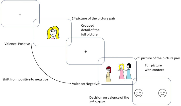
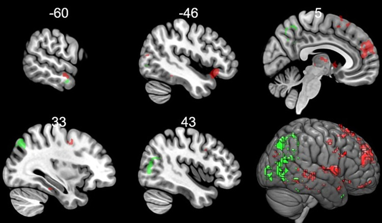
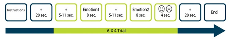
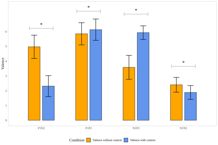
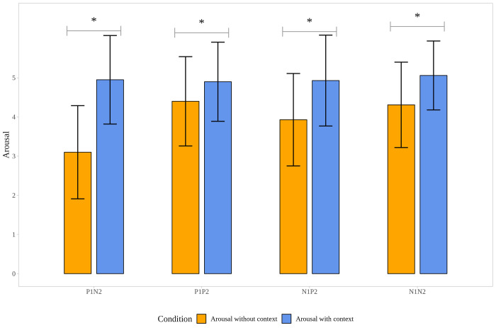
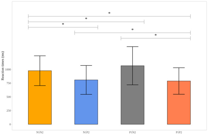
Emotional flexibility reflects the ability to adjust the emotional response to the changing environmental context. To understand how context can trigger a change in emotional response, i.e., how it can upregulate the initial emotional response or trigger a shift in the valence of emotional response, we used a task consisting of picture pairs during functional magnetic resonance imaging sessions. In each pair, the first picture was a smaller detail (a decontextualized photograph depicting emotions using primarily facial and postural expressions) from the second (contextualized) picture, and the neural response to a decontextualized picture was compared with the same picture in a context. Thirty-one healthy participants (18 females; mean age: 24.44 ± 3.4) were involved in the study. In general, context (vs. pictures without context) increased activation in areas involved in facial emotional processing (e.g., middle temporal gyrus, fusiform gyrus, and temporal pole) and affective mentalizing (e.g., precuneus, temporoparietal junction). After excluding the general effect of context by using an exclusive mask with activation to context vs. no-context, the automatic shift from positive to negative valence induced by the context was associated with increased activation in the thalamus, caudate, medial frontal gyrus and lateral orbitofrontal cortex. When the meaning changed from negative to positive, it resulted in a less widespread activation pattern, mainly in the precuneus, middle temporal gyrus, and occipital lobe. Providing context cues to facial information recruited brain areas that induced changes in the emotional responses and interpretation of the emotional situations automatically to support emotional flexibility.
情绪灵活性反映了根据环境变化调整情绪反应的能力。为了了解环境如何引发情绪反应的变化,即它如何上调初始情绪反应或引发情绪反应效价的转变,我们在功能磁共振成像期间使用了由图片对组成的任务。在每对中,第一张图片是第二张(上下文)图片的较小细节(使用主要面部和姿势表情描绘情绪的去语境化照片),并且比较了去语境化图片的神经反应与同一图片在上下文中的反应。 31 名健康参与者(18 名女性;平均年龄:24.44±3.4)参与了这项研究。一般来说,上下文(与没有上下文的图片相比)增加了参与面部情绪处理的区域(例如,颞中回、梭状回和颞极)和情感心理化(例如,楔前叶、颞顶联合区)的激活。通过使用仅对上下文与无上下文的激活进行对比的专用掩模排除上下文的一般影响后,上下文引起的从正性到负性效价的自动转变与丘脑、尾状核、内侧额回和外侧眶额皮层的激活增加有关。当意义从负性变为正性时,它导致了一种不那么广泛的激活模式,主要在楔前叶、颞中回和枕叶。为面部信息提供上下文线索会招募大脑区域,这些区域会自动引起情绪反应的变化和对情绪情境的解释,以支持情绪灵活性。