Department of Orthopaedics and Trauma Surgery, Musculoskeletal University Center Munich (MUM), University Hospital, LMU, Marchioninistr. 15, 81377, Munich, Germany.
Statistical Consulting Unit StaBLab, LMU, Munich, Germany.
Knee Surg Sports Traumatol Arthrosc. 2023 Apr;31(4):1483-1490. doi: 10.1007/s00167-022-07302-x. Epub 2023 Jan 3.
Many radiographic lower limb alignment measurements are dependent on patients' position, which makes a standardised image acquisition of long-leg radiographs (LLRs) essential for valid measurements. The purpose of this study was to investigate the influence of rotation and flexion of the lower limb on common radiological alignment parameters using three-dimensional (3D) simulation.
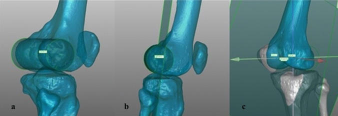
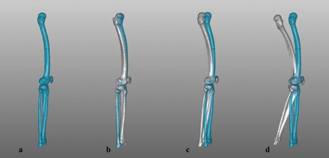
Joint angles and alignment parameters of 3D lower limb bone models (n = 60), generated from computed tomography (CT) scans, were assessed and projected into the coronal plane to mimic radiographic imaging. Bone models were subsequently rotated around the longitudinal mechanical axis up to 15° inward/outward and additionally flexed along the femoral intercondylar axis up to 30°. This resulted in 28 combinations of rotation and flexion for each leg. The results were statistically analysed on a descriptive level and using a linear mixed effects model.
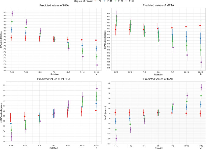
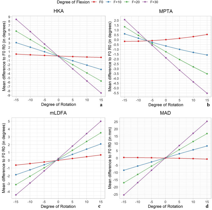
A total of 1680 simulations were performed. Mechanical axis deviation (MAD) revealed a medial deviation with increasing internal rotation and a lateral deviation with increasing external rotation. This effect increased significantly (p < 0.05) with combined flexion up to 30° flexion (- 25.4 mm to 25.2 mm). With the knee extended, the mean deviation of hip-knee-ankle angle (HKA) was small over all rotational steps but increased toward more varus/valgus when combined with flexion (8.4° to - 8.5°). Rotation alone changed the medial proximal tibial angle (MPTA) and the mechanical lateral distal femoral angle (mLDFA) in opposite directions, and the effects increased significantly (p < 0.05) when flexion was present.
Axial rotation and flexion of the 3D lower limb has a huge impact on the projected two-dimensional alignment measurements in the coronal plane. The observed effects were small for isolated rotation or flexion, but became pronounced and clinically relevant when there was a combination of both. This must be considered when evaluating X-ray images. Extension deficits of the knee make LLR prone to error and this calls into question direct postoperative alignment controls.
III (retrospective cohort study).
许多下肢放射学对线测量依赖于患者的体位,这使得长肢 X 线片(LLR)的标准图像采集对于有效测量至关重要。本研究的目的是使用三维(3D)模拟研究下肢旋转和弯曲对线参数的影响。
从计算机断层扫描(CT)扫描中生成 3D 下肢骨骼模型(n=60)的关节角度和对线参数,并将其投射到冠状面以模拟放射成像。随后,骨骼模型围绕纵向力学轴向内/向外旋转高达 15°,并沿股骨髁间轴弯曲高达 30°。这导致每条腿的旋转和弯曲有 28 种组合。结果在描述性水平和线性混合效应模型上进行了统计分析。
共进行了 1680 次模拟。机械轴偏差(MAD)显示出随着内旋的增加而向内侧偏斜,随着外旋的增加而向外侧偏斜。这种影响随着联合弯曲达到 30°弯曲而显著增加(p<0.05)(-25.4 毫米至 25.2 毫米)。当膝关节伸直时,所有旋转步骤中髋关节-膝关节-踝关节角度(HKA)的平均偏差都很小,但当与弯曲结合时,偏差会向更内翻/外翻增加(8.4°至-8.5°)。旋转本身会使内侧近端胫骨角(MPTA)和机械外侧远端股骨角(mLDFA)朝相反方向改变,当存在弯曲时,这些影响显著增加(p<0.05)。
3D 下肢的轴向旋转和弯曲对线在冠状面上的二维对线测量有很大影响。单独旋转或弯曲的影响较小,但当两者结合时,影响明显且具有临床相关性。这在评估 X 射线图像时必须考虑。膝关节伸展不足使 LLR 容易出现误差,这对直接术后对线控制提出了质疑。
III(回顾性队列研究)。