Zhang Xuede, Bao Lingling, Yu Guohua, Wang Haifeng
Department of Oncology, Weifang People's Hospital, Weifang, China.
Department of Hematology and Oncology, Beilun District People's Hospital, Ningbo, China.
Front Surg. 2023 Jan 6;9:1050242. doi: 10.3389/fsurg.2022.1050242. eCollection 2022.
Pleural effusion (PE) caused by lung cancer is prevalent, and it is difficult to differentiate it from PE caused by tuberculosis. Exosome-based liquid biopsy offers a non-invasive technique to diagnose benign and malignant PE. Exosomal miRNAs are potential diagnostic markers and play an essential role in signal transduction and biological processes in tumor development. We hypothesized that exosomal miRNA expression profiles in PE would contribute to identifying its diagnostic markers and elucidating the molecular basis of PE formation in lung cancer.
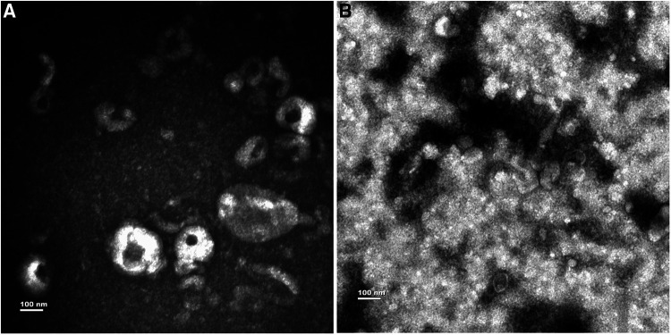
The exosomes from PE caused by lung adenocarcinoma (LUAD) and pulmonary tuberculosis were isolated and verified by transmission electron microscopy. The exosomal miRNA profiles were identified using deep sequencing and validated with quantitative real-time PCR (qRT-PCR). We performed bioinformatic analysis for differentially expressed miRNAs to explore how exosomal miRNAs regulate pleural effusion.
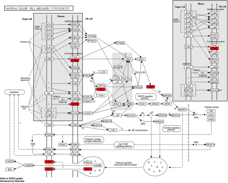
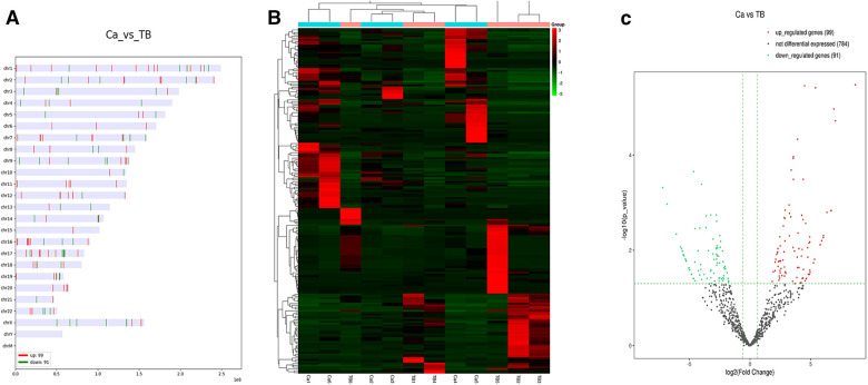
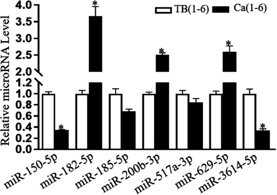
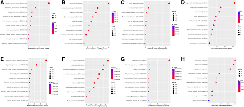
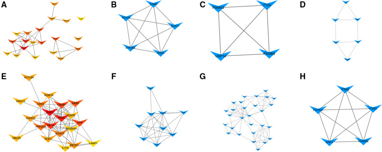
We identified 99 upregulated and 91 downregulated miRNAs in malignant pleural effusion (MPE) compared to tuberculous pleural effusion (TPE). Seven differentially expressed miRNAs (DEmiRNAs) were validated by qRT-PCR, out of which 5 (71.4%) were confirmed through sequencing. Gene Ontology (GO) analysis revealed that most exosomal miRNAs target genes were involved in regulating cellular processes and nitrogen compound metabolism. According to the Kyoto Encyclopedia of Genes and Genomes (KEGG) pathway enrichment analysis, the exosomal miRNAs target genes were mainly involved in Fc gamma R-mediated phagocytosis, Rap1 signaling pathway, and breast cancer. The hub genes, including ITGAM, FOXO1, MAPK14, YWHAB, GRIN1, and PRF1, were screened through plug-in cytoHubba. The PFR1 was identified as a critical gene in MPE formation using single-cell sequencing analysis. Additionally, we hypothesized that tumor cells affected natural killer cells and promoted the generation of PE in LUAD the exosomal hsa-miR-3120-5p-PRF1 axis.
We identified exosomal miRNA profiles in LUAD-MPE and TPE, which may help in the differential diagnosis of MPE and TPE. Bioinformatic analysis revealed that these miRNAs might affect PE generation through tumor immune response in LUAD. Our results provided a new theoretical basis for understanding the function of exosomal miRNAs in LUAD-MPE.
肺癌所致胸腔积液(PE)较为常见,且难以与结核性胸腔积液相鉴别。基于外泌体的液体活检提供了一种非侵入性技术用于诊断良性和恶性胸腔积液。外泌体微小RNA(miRNA)是潜在的诊断标志物,在肿瘤发生发展的信号转导和生物学过程中发挥重要作用。我们推测,胸腔积液中外泌体miRNA表达谱有助于识别其诊断标志物并阐明肺癌中胸腔积液形成的分子基础。
分离并通过透射电子显微镜验证了肺腺癌(LUAD)和肺结核所致胸腔积液中的外泌体。使用深度测序鉴定外泌体miRNA谱,并通过定量实时聚合酶链反应(qRT-PCR)进行验证。我们对差异表达的miRNA进行生物信息学分析,以探讨外泌体miRNA如何调节胸腔积液。
与结核性胸腔积液(TPE)相比,我们在恶性胸腔积液(MPE)中鉴定出99个上调的miRNA和91个下调的miRNA。通过qRT-PCR验证了7个差异表达的miRNA(DEmiRNAs),其中5个(71.4%)通过测序得到确认。基因本体(GO)分析显示,大多数外泌体miRNA靶基因参与调节细胞过程和氮化合物代谢。根据京都基因与基因组百科全书(KEGG)通路富集分析,外泌体miRNA靶基因主要参与FcγR介导的吞噬作用、Rap1信号通路和乳腺癌。通过插件cytoHubba筛选出包括整合素αM(ITGAM)、叉头框蛋白O1(FOXO1)、丝裂原活化蛋白激酶14(MAPK14)、14-3-3蛋白β/α(YWHAB)、谷氨酸离子型受体NMDA 1型亚基(GRIN1)和穿孔素1(PRF1)在内的枢纽基因。通过单细胞测序分析确定PFR1是MPE形成中的关键基因。此外,我们推测肿瘤细胞影响自然杀伤细胞并通过外泌体hsa-miR-3120-5p-PRF1轴促进LUAD中胸腔积液的产生。
我们鉴定了LUAD-MPE和TPE中的外泌体miRNA谱,这可能有助于MPE和TPE的鉴别诊断。生物信息学分析表明,这些miRNA可能通过LUAD中的肿瘤免疫反应影响胸腔积液的产生。我们的结果为理解外泌体miRNA在LUAD-MPE中的功能提供了新的理论基础。