Wong Daniel R, Magaki Shino D, Vinters Harry V, Yong William H, Monuki Edwin S, Williams Christopher K, Martini Alessandra C, DeCarli Charles, Khacherian Chris, Graff John P, Dugger Brittany N, Keiser Michael J
Institute for Neurodegenerative Diseases, University of California, San Francisco, San Francisco, CA, 94158, USA.
Bakar Computational Health Sciences Institute, University of California, San Francisco, CA, 94158, USA.
bioRxiv. 2023 Jan 17:2023.01.13.524019. doi: 10.1101/2023.01.13.524019.
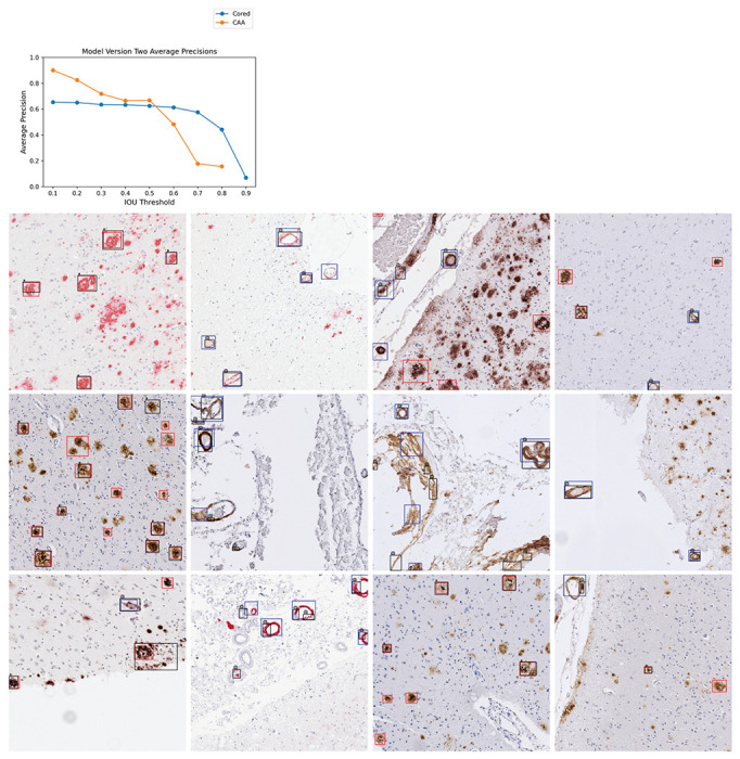
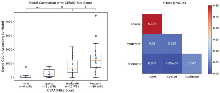
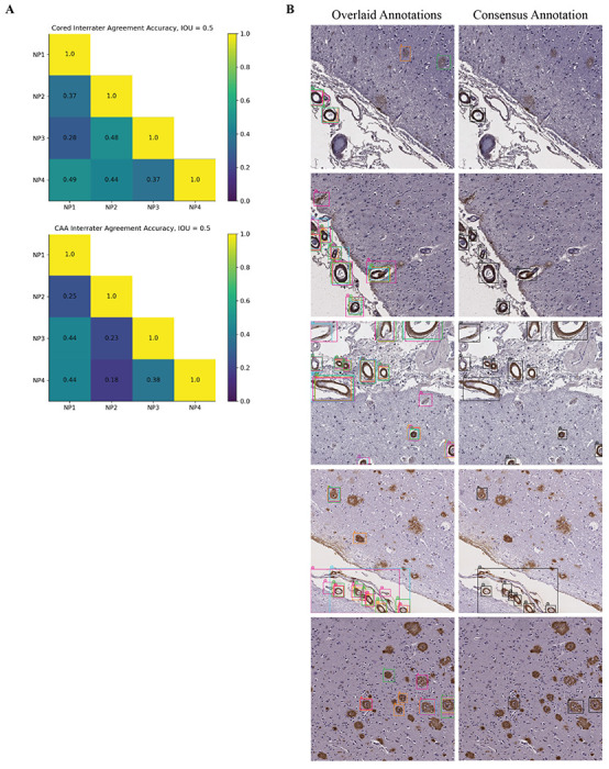
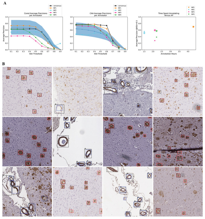
Precise, scalable, and quantitative evaluation of whole slide images is crucial in neuropathology. We release a deep learning model for rapid object detection and precise information on the identification, locality, and counts of cored plaques and cerebral amyloid angiopathies (CAAs). We trained this object detector using a repurposed image-tile dataset without any human-drawn bounding boxes. We evaluated the detector on a new manually-annotated dataset of whole slide images (WSIs) from three institutions, four staining procedures, and four human experts. The detector matched the cohort of neuropathology experts, achieving 0.64 (model) vs. 0.64 (cohort) average precision (AP) for cored plaques and 0.75 vs. 0.51 AP for CAAs at a 0.5 IOU threshold. It provided count and locality predictions that correlated with gold-standard CERAD-like WSI scoring (p=0.07± 0.10). The openly-available model can quickly score WSIs in minutes without a GPU on a standard workstation.
在神经病理学中,对全玻片图像进行精确、可扩展且定量的评估至关重要。我们发布了一个深度学习模型,用于快速目标检测以及获取有关核心斑块和脑淀粉样血管病(CAA)的识别、定位和计数的精确信息。我们使用一个重新利用的图像块数据集训练了这个目标检测器,该数据集中没有任何人工绘制的边界框。我们在一个来自三个机构、四种染色程序以及四位人类专家的新的全玻片图像(WSI)人工标注数据集上对该检测器进行了评估。在0.5的交并比(IOU)阈值下,该检测器与神经病理学专家团队的表现相当,核心斑块的平均精度(AP)为0.64(模型)对0.64(专家团队),CAA的平均精度为0.75对0.51。它提供的计数和定位预测与金标准的类CERAD的WSI评分相关(p = 0.07±0.10)。这个公开可用的模型在标准工作站上无需GPU即可在几分钟内快速对WSI进行评分。