Department of Oral and Maxillofacial Surgery, University Hospital of RWTH Aachen, Pauwelsstraße 30, 52074, Aachen, Germany.
Department of Orthopedics, Trauma and Reconstructive Surgery, University Hospital of RWTH Aachen, Pauwelsstraße 30, 52074, Aachen, Germany.
BMC Oral Health. 2023 Jan 31;23(1):56. doi: 10.1186/s12903-023-02726-4.
A rigorous search for alternatives to autogenous bone grafts to avoid invasiveness at the donor site in the treatment of maxillomandibular bone defects. Researchers have used alloplastic, allogeneic, and xenogeneic bone graft substitutes in clinical studies with varying degrees of success, although their in vitro effects on stem cells remain unclear. Dental pulp stem cells (DPSCs) can potentially enhance the bone regeneration of bone graft substitutes. The present in vitro study investigates the osteogenic capability of DPSCs on alloplastic (biphasic calcium phosphate [BCP]), allogeneic (freeze-dried bone allografts [FDBAs]), and xenogeneic (deproteinized bovine bone mineral [DBBM]) bone grafts.
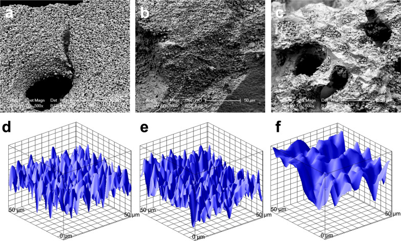
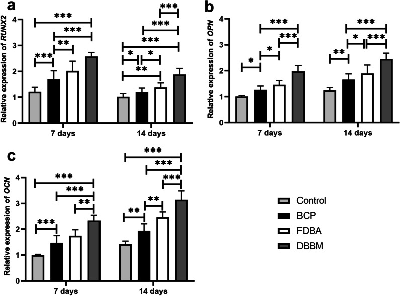
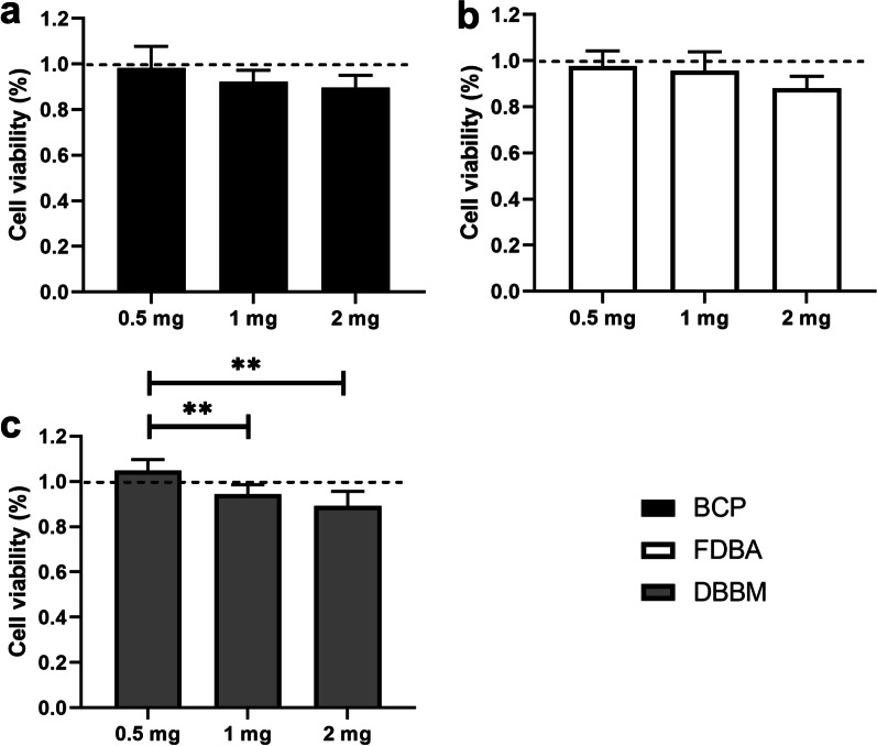
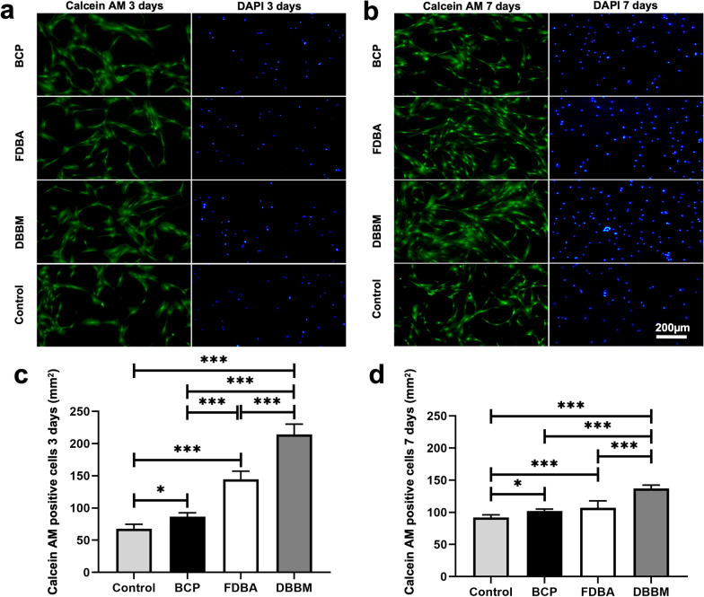
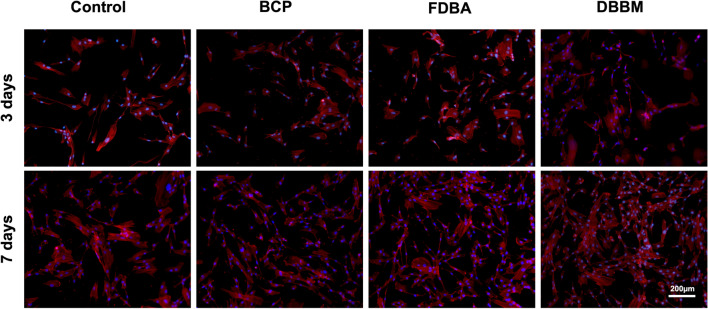
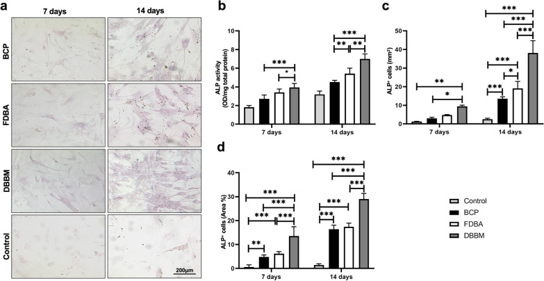
Human DPSCs were seeded on 0.5 mg/ml, 1 mg/ml, and 2 mg/ml of BCP, FDBA, and DBBM to evaluate the optimal cell growth and cytotoxicity. Scaffolds and cell morphologies were analyzed by scanning electron microscopy (SEM). Calcein AM and cytoskeleton staining were performed to determine cell attachment and proliferation. Alkaline phosphatase (ALP) and osteogenesis-related genes expressions was used to investigate initial osteogenic differentiation.
Cytotoxicity assays showed that most viable DPSCs were present at a scaffold concentration of 0.5 mg/ml. The DPSCs on the DBBM scaffold demonstrated a significantly higher proliferation rate of 214.25 ± 16.17 (p < 0.001) cells, enhancing ALP activity level and upregulating of osteogenesis-related genes compared with other two scaffolds.
DBBP scaffold led to extremely high cell viability, but also promoted proliferation, attachment, and enhanced the osteogenic differentiation capacity of DPSCs, which hold great potential for bone regeneration treatment; however, further studies are necessary.
为了避免在治疗上颌下颌骨缺损时在供体部位产生侵入性,研究人员一直在寻找替代自体骨移植物的方法。在临床研究中,研究人员已经使用了同种异体、异种和合成骨移植替代物,取得了不同程度的成功,尽管它们对干细胞的体外影响尚不清楚。牙髓干细胞(DPSCs)有可能增强骨移植物替代物的骨再生能力。本体外研究调查了 DPSCs 对同种异体(双相磷酸钙[BCP])、同种异体(冻干骨同种异体[FDBAs])和异种(去蛋白牛骨矿物质[DBBM])骨移植物的成骨能力。
将人牙髓干细胞接种于 0.5mg/ml、1mg/ml 和 2mg/ml 的 BCP、FDBA 和 DBBM 上,以评估最佳细胞生长和细胞毒性。通过扫描电子显微镜(SEM)分析支架和细胞形态。通过钙黄绿素 AM 和细胞骨架染色来确定细胞附着和增殖。碱性磷酸酶(ALP)和骨生成相关基因表达用于研究初始成骨分化。
细胞毒性试验表明,在支架浓度为 0.5mg/ml 时,大多数活的 DPSCs 存在。与其他两种支架相比,DBBM 支架上的 DPSCs 增殖率显著更高,为 214.25±16.17(p<0.001)细胞,增强了 ALP 活性水平和上调了骨生成相关基因。
DBBP 支架导致细胞活力极高,但也促进了 DPSCs 的增殖、附着和增强了其成骨分化能力,这为骨再生治疗提供了巨大的潜力;然而,还需要进一步的研究。