• 文献检索
  • 文档翻译
  • 深度研究
  • 学术资讯
  • Suppr Zotero 插件Zotero 插件
  • 邀请有礼
  • 套餐&价格
  • 历史记录
应用&插件
Suppr Zotero 插件Zotero 插件浏览器插件Mac 客户端Windows 客户端微信小程序
定价
高级版会员购买积分包购买API积分包
服务
文献检索文档翻译深度研究API 文档MCP 服务
关于我们
关于 Suppr公司介绍联系我们用户协议隐私条款
关注我们

Suppr 超能文献

核心技术专利:CN118964589B侵权必究
粤ICP备2023148730 号-1Suppr @ 2026

文献检索

告别复杂PubMed语法,用中文像聊天一样搜索,搜遍4000万医学文献。AI智能推荐,让科研检索更轻松。

立即免费搜索

文件翻译

保留排版,准确专业,支持PDF/Word/PPT等文件格式,支持 12+语言互译。

免费翻译文档

深度研究

AI帮你快速写综述,25分钟生成高质量综述,智能提取关键信息,辅助科研写作。

立即免费体验

利用单细胞分析技术创建口腔炎症相关骨病骨微环境图谱。

Creating an atlas of the bone microenvironment during oral inflammatory-related bone disease using single-cell profiling.

机构信息

State Key Laboratory of Oral Diseases, National Clinical Research Center for Oral Diseases, Department of Cariology and Endodontics, West China Hospital of Stomatology, Sichuan University, Chengdu, China.

State Key Laboratory of Oral Diseases, National Clinical Research Center for Oral Diseases, Department of Orthognathic and TMJ Surgery, West China Hospital of Stomatology, Sichuan University, Chengdu, China.

出版信息

Elife. 2023 Feb 1;12:e82537. doi: 10.7554/eLife.82537.

DOI:10.7554/eLife.82537
PMID:36722472
原文链接:https://pmc.ncbi.nlm.nih.gov/articles/PMC9925051/
Abstract

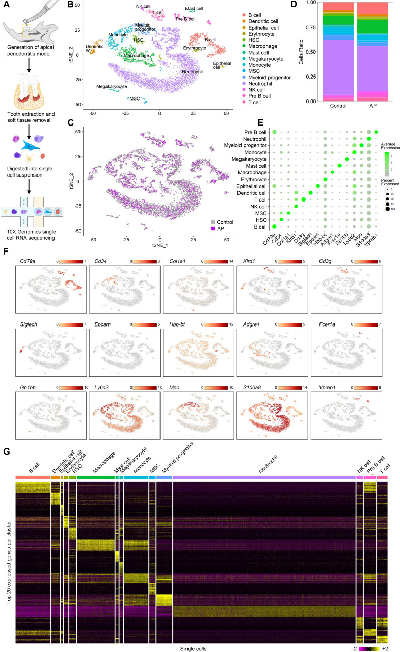
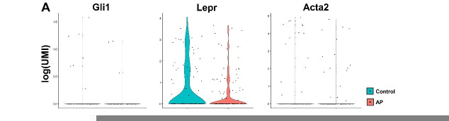
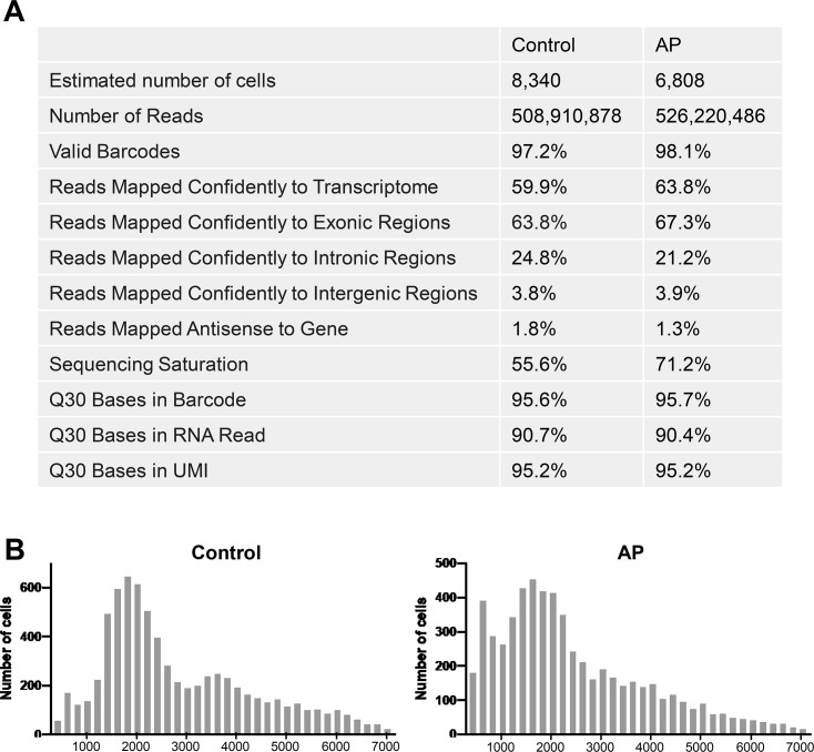
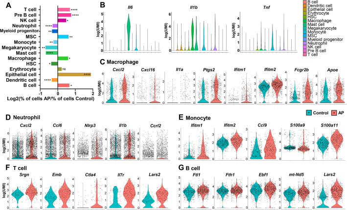
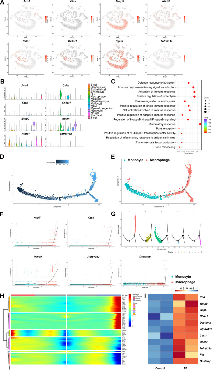
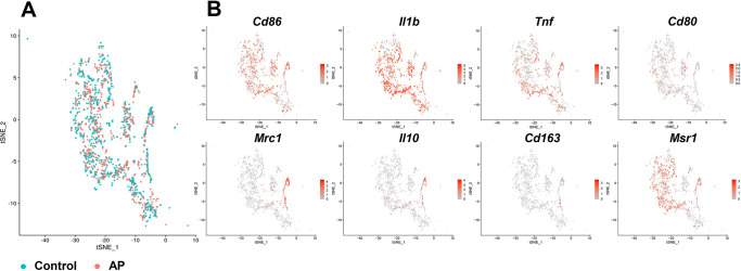
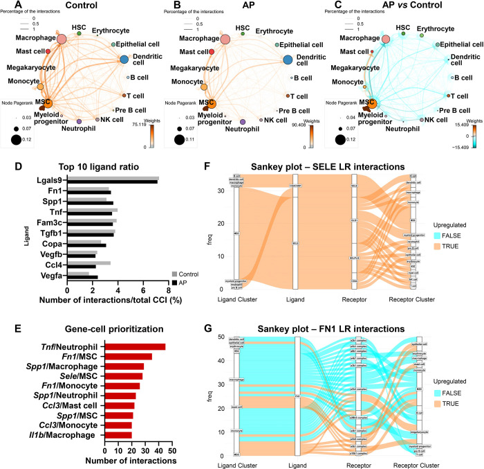
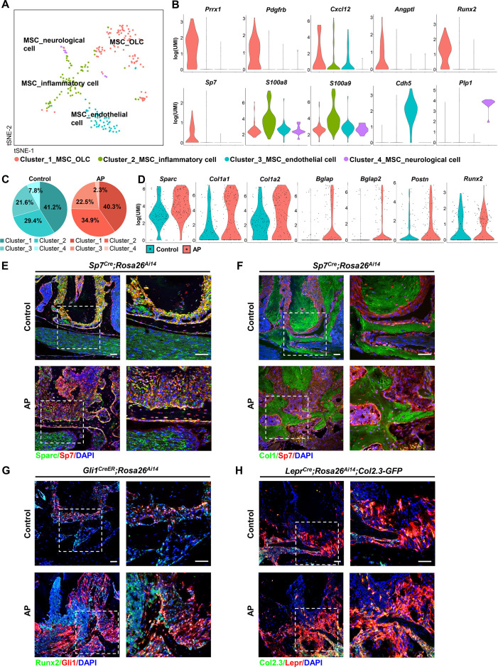
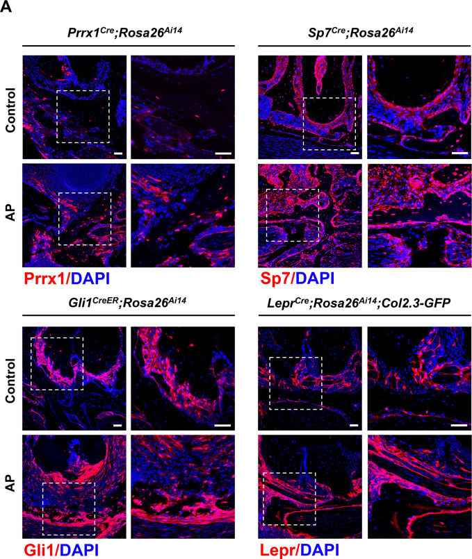
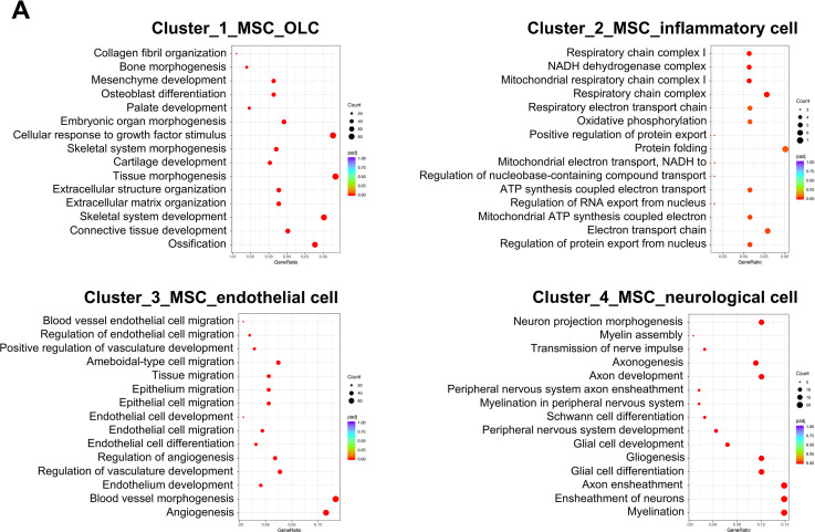
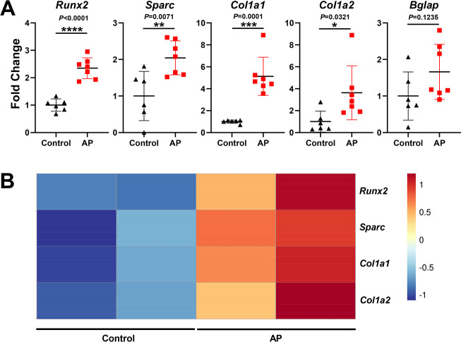
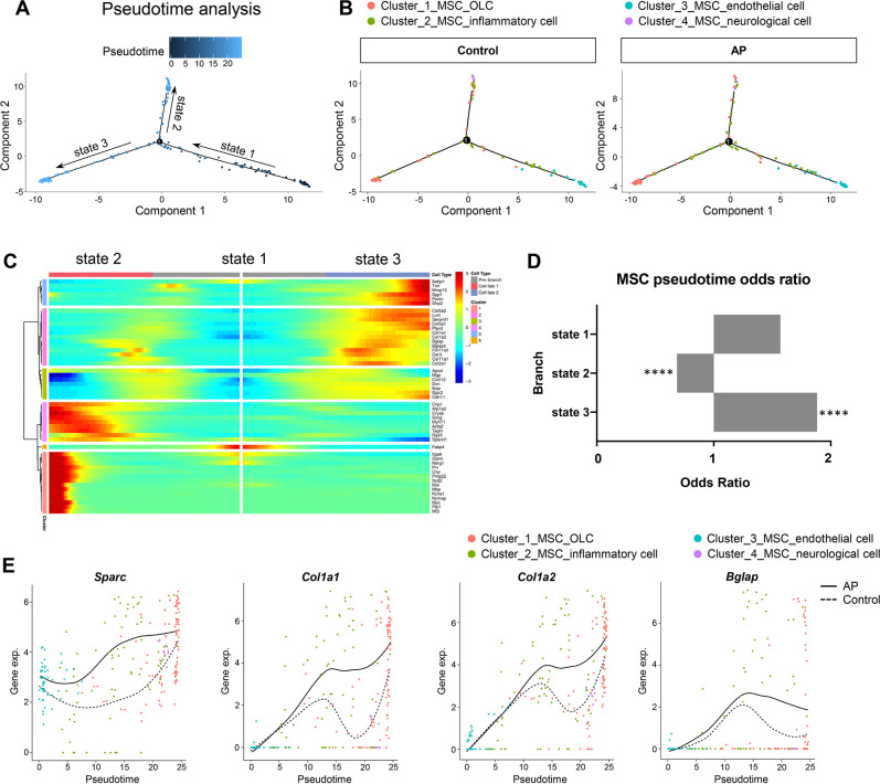
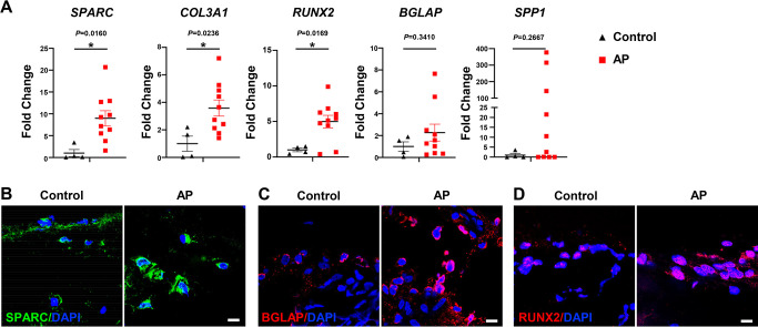
Oral inflammatory diseases such as apical periodontitis are common bacterial infectious diseases that may affect the periapical alveolar bone tissues. A protective process occurs simultaneously with the inflammatory tissue destruction, in which mesenchymal stem cells (MSCs) play a primary role. However, a systematic and precise description of the cellular and molecular composition of the microenvironment of bone affected by inflammation is lacking. In this study, we created a single-cell atlas of cell populations that compose alveolar bone in healthy and inflammatory disease states. We investigated changes in expression frequency and patterns related to apical periodontitis, as well as the interactions between MSCs and immunocytes. Our results highlight an enhanced self-supporting network and osteogenic potential within MSCs during apical periodontitis-associated inflammation. MSCs not only differentiated toward osteoblast lineage cells but also expressed higher levels of osteogenic-related markers, including Sparc and Col1a1. This was confirmed by lineage tracing in transgenic mouse models and human samples from oral inflammatory-related alveolar bone lesions. In summary, the current study provides an in-depth description of the microenvironment of MSCs and immunocytes in both healthy and disease states. We also identified key apical periodontitis-associated MSC subclusters and their biomarkers, which could further our understanding of the protective process and the underlying mechanisms of oral inflammatory-related bone disease. Taken together, these results enhance our understanding of heterogeneity and cellular interactions of alveolar bone cells under pathogenic and inflammatory conditions. We provide these data as a tool for investigators not only to better appreciate the repertoire of progenitors that are stress responsive but importantly to help design new therapeutic targets to restore bone lesions caused by apical periodontitis and other inflammatory-related bone diseases.

摘要

口腔炎症性疾病,如根尖周炎,是常见的细菌性传染病,可能影响根尖牙槽骨组织。在炎症组织破坏的同时,会发生保护过程,间充质干细胞(MSCs)在其中发挥主要作用。然而,对于受炎症影响的骨微环境的细胞和分子组成,缺乏系统和精确的描述。在这项研究中,我们创建了一个单细胞图谱,描绘了健康和炎症疾病状态下组成牙槽骨的细胞群体。我们研究了与根尖周炎相关的表达频率和模式的变化,以及 MSCs 和免疫细胞之间的相互作用。我们的结果突出了在根尖周炎相关炎症期间,MSCs 内部增强的自我支持网络和成骨潜能。MSCs 不仅向成骨细胞谱系细胞分化,而且表达更高水平的成骨相关标志物,包括 Sparc 和 Col1a1。这一点通过转基因小鼠模型和口腔炎症相关牙槽骨病变的人类样本中的谱系追踪得到了证实。总之,本研究深入描述了健康和疾病状态下 MSCs 和免疫细胞的微环境。我们还确定了与根尖周炎相关的 MSC 亚群及其生物标志物,这可能有助于我们深入了解保护性过程和与口腔炎症相关的骨疾病的潜在机制。综上所述,这些结果增强了我们对致病和炎症条件下牙槽骨细胞异质性和细胞相互作用的理解。我们提供这些数据不仅是为了让研究人员更好地了解对压力有反应的祖细胞的多样性,而且更重要的是帮助设计新的治疗靶点,以恢复根尖周炎和其他与炎症相关的骨疾病引起的骨损伤。

https://cdn.ncbi.nlm.nih.gov/pmc/blobs/9d61/9925051/5daa60f5c010/elife-82537-sa2-fig1.jpg
https://cdn.ncbi.nlm.nih.gov/pmc/blobs/9d61/9925051/c610efc16731/elife-82537-fig1.jpg
https://cdn.ncbi.nlm.nih.gov/pmc/blobs/9d61/9925051/985920b6b1e0/elife-82537-fig1-figsupp1.jpg
https://cdn.ncbi.nlm.nih.gov/pmc/blobs/9d61/9925051/76d662c0433f/elife-82537-fig2.jpg
https://cdn.ncbi.nlm.nih.gov/pmc/blobs/9d61/9925051/4d13f8e02336/elife-82537-fig3.jpg
https://cdn.ncbi.nlm.nih.gov/pmc/blobs/9d61/9925051/c7dc7f7904b4/elife-82537-fig3-figsupp1.jpg
https://cdn.ncbi.nlm.nih.gov/pmc/blobs/9d61/9925051/adc8be50fe15/elife-82537-fig4.jpg
https://cdn.ncbi.nlm.nih.gov/pmc/blobs/9d61/9925051/70cd34f547e9/elife-82537-fig5.jpg
https://cdn.ncbi.nlm.nih.gov/pmc/blobs/9d61/9925051/cddb4f22126d/elife-82537-fig5-figsupp1.jpg
https://cdn.ncbi.nlm.nih.gov/pmc/blobs/9d61/9925051/014e3ca48941/elife-82537-fig5-figsupp2.jpg
https://cdn.ncbi.nlm.nih.gov/pmc/blobs/9d61/9925051/2f1f9ebc0f22/elife-82537-fig5-figsupp3.jpg
https://cdn.ncbi.nlm.nih.gov/pmc/blobs/9d61/9925051/bf2ec7976333/elife-82537-fig6.jpg
https://cdn.ncbi.nlm.nih.gov/pmc/blobs/9d61/9925051/4eebf04601e1/elife-82537-fig7.jpg
https://cdn.ncbi.nlm.nih.gov/pmc/blobs/9d61/9925051/5daa60f5c010/elife-82537-sa2-fig1.jpg

相似文献

1
Creating an atlas of the bone microenvironment during oral inflammatory-related bone disease using single-cell profiling.利用单细胞分析技术创建口腔炎症相关骨病骨微环境图谱。
Elife. 2023 Feb 1;12:e82537. doi: 10.7554/eLife.82537.
2
Protective Actions in Apical Periodontitis: The Regenerative Bioactivities Led by Mesenchymal Stem Cells.根尖周病的防护措施:间充质干细胞的再生生物活性。
Biomolecules. 2022 Nov 23;12(12):1737. doi: 10.3390/biom12121737.
3
Dickkopf-1 may regulate bone coupling by attenuating wnt/β-catenin signaling in chronic apical periodontitis.Dickkopf-1 可能通过减弱慢性根尖周炎中的 Wnt/β-连环蛋白信号来调节骨偶联。
Arch Oral Biol. 2018 Feb;86:94-100. doi: 10.1016/j.archoralbio.2017.11.012. Epub 2017 Dec 5.
4
Aging-Affected MSC Functions and Severity of Periodontal Tissue Destruction in a Ligature-Induced Mouse Periodontitis Model.衰老对间充质干细胞功能的影响及结扎诱导的小鼠牙周炎模型中牙周组织破坏的严重程度。
Int J Mol Sci. 2020 Oct 30;21(21):8103. doi: 10.3390/ijms21218103.
5
MiR-17 modulates osteogenic differentiation through a coherent feed-forward loop in mesenchymal stem cells isolated from periodontal ligaments of patients with periodontitis.miR-17 通过整合反馈环调控牙周炎患者牙周膜间充质干细胞的成骨分化。
Stem Cells. 2011 Nov;29(11):1804-16. doi: 10.1002/stem.728.
6
Notch signaling pathway mediates alveolar bone resorption in apical periodontitis.Notch 信号通路介导根尖周炎中的牙槽骨吸收。
Med Hypotheses. 2019 Mar;124:87-90. doi: 10.1016/j.mehy.2019.02.018. Epub 2019 Feb 4.
7
NLRP12 Attenuates Inflammatory Bone Loss in Experimental Apical Periodontitis.NLRP12 减轻实验性根尖周炎的炎症性骨丢失。
J Dent Res. 2019 Apr;98(4):476-484. doi: 10.1177/0022034518820289. Epub 2019 Jan 25.
8
Role of the Btk-PLC2 Signaling Pathway in the Bone Destruction of Apical Periodontitis.BTK-PLC2 信号通路在根尖周炎骨破坏中的作用。
Mediators Inflamm. 2019 Jul 25;2019:8767529. doi: 10.1155/2019/8767529. eCollection 2019.
9
Single-cell RNA landscape of the osteoimmunology microenvironment in periodontitis.单细胞 RNA 图谱揭示牙周炎中的骨免疫学微环境。
Theranostics. 2022 Jan 1;12(3):1074-1096. doi: 10.7150/thno.65694. eCollection 2022.
10
The possible role of Gremlin1 in inflammatory apical periodontitis.Gremlin1 在炎症性根尖周炎中的可能作用。
Arch Oral Biol. 2024 Jan;157:105848. doi: 10.1016/j.archoralbio.2023.105848. Epub 2023 Nov 10.

引用本文的文献

1
The inhibitory effects of 7ND protein on osteoclast differentiation in apical periodontitis.7ND蛋白对根尖周炎中破骨细胞分化的抑制作用。
Front Cell Infect Microbiol. 2025 Jun 27;15:1597604. doi: 10.3389/fcimb.2025.1597604. eCollection 2025.
2
The Oral Microbiome and Systemic Health: Bridging the Gap Between Dentistry and Medicine.口腔微生物群与全身健康:弥合牙科与医学之间的差距。
Cureus. 2025 Feb 12;17(2):e78918. doi: 10.7759/cureus.78918. eCollection 2025 Feb.
3
The heterogeneity of T cell infiltration in human periapical lesions.

本文引用的文献

1
Protective Actions in Apical Periodontitis: The Regenerative Bioactivities Led by Mesenchymal Stem Cells.根尖周病的防护措施:间充质干细胞的再生生物活性。
Biomolecules. 2022 Nov 23;12(12):1737. doi: 10.3390/biom12121737.
2
Current Developments in the Preclinical and Clinical use of Natural Killer T cells.自然杀伤 T 细胞的临床前和临床应用的最新进展。
BioDrugs. 2023 Jan;37(1):57-71. doi: 10.1007/s40259-022-00572-4. Epub 2022 Dec 16.
3
Macrophages in periapical lesions: Potential roles and future directions.根尖周病变中的巨噬细胞:潜在作用和未来方向。
人类根尖周病变中T细胞浸润的异质性。
BMC Oral Health. 2025 Feb 19;25(1):261. doi: 10.1186/s12903-025-05621-2.
4
Chemokine receptor-2 deficiency induced mild experimental periapical lesion in mice.趋化因子受体-2缺陷在小鼠中诱发轻度实验性根尖周病变。
J Dent Sci. 2025 Jan;20(1):402-409. doi: 10.1016/j.jds.2024.06.005. Epub 2024 Jun 14.
5
Single-Cell and Transcriptome Analysis of Periodontitis: Molecular Subtypes and Biomarkers Linked to Mitochondrial Dysfunction and Immunity.牙周炎的单细胞和转录组分析:与线粒体功能障碍和免疫相关的分子亚型及生物标志物
J Inflamm Res. 2024 Dec 27;17:11659-11678. doi: 10.2147/JIR.S498739. eCollection 2024.
6
Negative feedback between PTH1R and IGF1 through the Hedgehog pathway in mediating craniofacial bone remodeling.甲状旁腺激素1型受体(PTH1R)与胰岛素样生长因子1(IGF1)之间通过刺猬信号通路产生的负反馈在介导颅面骨重塑过程中发挥作用。
JCI Insight. 2024 Dec 17;10(3):e183684. doi: 10.1172/jci.insight.183684.
7
Nanoparticles and bone microenvironment: a comprehensive review for malignant bone tumor diagnosis and treatment.纳米颗粒与骨微环境:恶性骨肿瘤诊治的全面综述
Mol Cancer. 2024 Nov 1;23(1):246. doi: 10.1186/s12943-024-02161-1.
8
Advances in hybridized nanoarchitectures for improved oro-dental health.用于改善口腔健康的杂交纳米结构的进展。
J Nanobiotechnology. 2024 Aug 7;22(1):469. doi: 10.1186/s12951-024-02680-5.
9
Periapical lesion-derived decellularized extracellular matrix as a potential solution for regenerative endodontics.根尖周病变来源的脱细胞细胞外基质作为再生牙髓病学的潜在解决方案。
Regen Biomater. 2024 May 7;11:rbae050. doi: 10.1093/rb/rbae050. eCollection 2024.
10
The neutrophil-osteogenic cell axis promotes bone destruction in periodontitis.中性粒细胞-成骨细胞轴促进牙周炎中的骨破坏。
Int J Oral Sci. 2024 Feb 27;16(1):18. doi: 10.1038/s41368-023-00275-8.
Front Immunol. 2022 Sep 5;13:949102. doi: 10.3389/fimmu.2022.949102. eCollection 2022.
4
Klotho in Osx-mesenchymal progenitors exerts pro-osteogenic and anti-inflammatory effects during mandibular alveolar bone formation and repair.Klotho 在 Osx-间充质祖细胞中发挥促成骨和抗炎作用,参与下颌牙槽骨形成和修复。
Signal Transduct Target Ther. 2022 May 11;7(1):155. doi: 10.1038/s41392-022-00957-5.
5
CrossTalkeR: analysis and visualization of ligand-receptorne tworks.CrossTalkeR:配体-受体网络的分析和可视化。
Bioinformatics. 2021 Nov 18;37(22):4263-4265. doi: 10.1093/bioinformatics/btab370.
6
The Potential Roles of T Cells in Periapical Lesions.T 细胞在根尖病变中的潜在作用。
J Endod. 2022 Jan;48(1):70-79. doi: 10.1016/j.joen.2021.09.016. Epub 2021 Oct 7.
7
B-Cell RANKL Contributes to Pathogen-Induced Alveolar Bone Loss in an Experimental Periodontitis Mouse Model.B细胞受体激活因子配体在实验性牙周炎小鼠模型中促成病原体诱导的牙槽骨丧失。
Front Physiol. 2021 Sep 14;12:722859. doi: 10.3389/fphys.2021.722859. eCollection 2021.
8
Mesenchymal Stem Cell-Macrophage Crosstalk and Maintenance of Inflammatory Microenvironment Homeostasis.间充质干细胞与巨噬细胞的相互作用及炎症微环境稳态的维持
Front Cell Dev Biol. 2021 Jun 25;9:681171. doi: 10.3389/fcell.2021.681171. eCollection 2021.
9
A single-cell atlas of human teeth.人类牙齿的单细胞图谱。
iScience. 2021 Apr 23;24(5):102405. doi: 10.1016/j.isci.2021.102405. eCollection 2021 May 21.
10
Mapping the immune microenvironment for mandibular alveolar bone homeostasis at single-cell resolution.以单细胞分辨率绘制下颌牙槽骨稳态的免疫微环境图谱。
Bone Res. 2021 Mar 15;9(1):17. doi: 10.1038/s41413-021-00141-5.