F. Joseph Halcomb III, M.D. Department of Biomedical Engineering, University of Kentucky, Lexington, Kentucky, United States of America.
Department of Microbiology, Immunology & Molecular Genetics, University of Kentucky, Lexington, Kentucky, United States of America.
PLoS One. 2023 Feb 2;18(2):e0280746. doi: 10.1371/journal.pone.0280746. eCollection 2023.
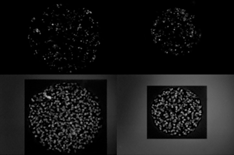
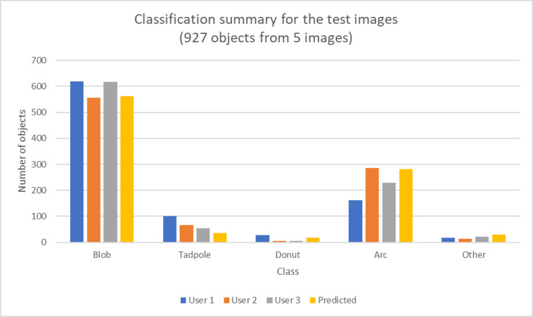
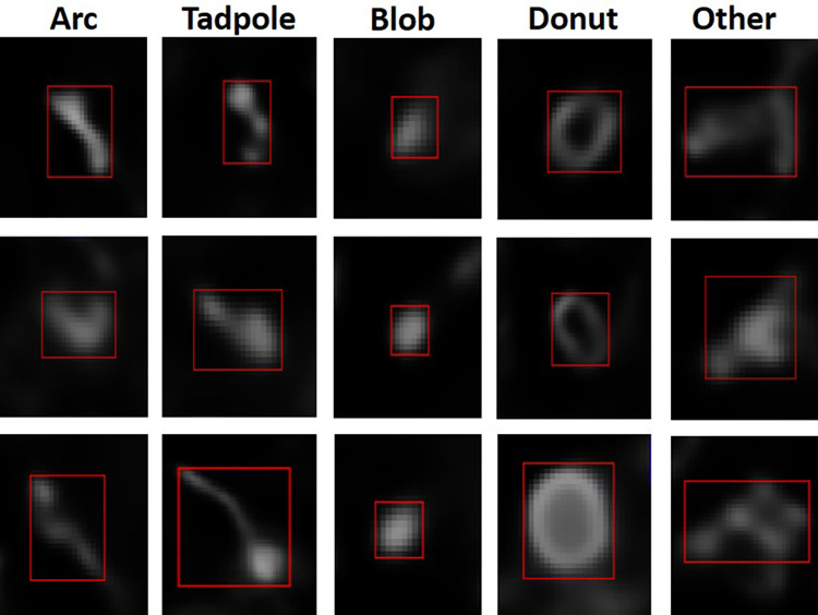
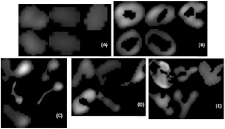
The mitochondrion is intimately linked to energy and overall metabolism and therefore the morphology of mitochondrion can be very informative for inferring the metabolic state of cells. In this study we report an approach for automatic classification of mitochondrial morphologies using supervised machine learning to efficiently classify them from a large number of cells at a time. Fluorescence microscopy images of the chronic encysted form of parasite Toxoplasma gondii were used for this development. Manually classifying these morphologies from the hundreds of parasites within typical tissue cysts is tedious and error prone. In addition, because of inherent biological heterogeneity in morphologies, there can be variability and lack of reproducibility in manual classification. We used image segmentation to detect mitochondrial shapes and used features extracted from them in a multivariate logistic regression model to classify the detected shapes into five morphological classes: Blobs, Tadpoles, Lasso/Donuts, Arcs, and Other. The detected shapes from a subset of images were first used to obtain consensus classification among expert users to obtain a labeled set. The model was trained using the labeled set from five cysts and its performance was tested on the mitochondrial morphologies from ten other cysts that were not used in training. Results showed that the model had an average overall accuracy of 87%. There was high degree of confidence in the classification of Blobs and Arcs (average F scores 0.91 and 0.73) which constituted the majority of morphologies (85%). Although the current development used microscopy images from tissue cysts of Toxoplasma gondii, the approach is adaptable with minor adjustments and can be used to automatically classify morphologies of organelles from a variety of cells.
线粒体与能量和整体代谢密切相关,因此线粒体的形态可以为推断细胞的代谢状态提供非常有价值的信息。在这项研究中,我们报告了一种使用监督机器学习自动分类线粒体形态的方法,以便能够一次从大量细胞中高效地对其进行分类。我们使用了慢性包囊型寄生虫弓形虫的荧光显微镜图像来进行这项开发。从典型的组织包囊中数百个寄生虫中手动对这些形态进行分类既繁琐又容易出错。此外,由于形态学上固有的生物学异质性,手动分类可能存在变异性和缺乏可重复性。我们使用图像分割来检测线粒体的形状,并使用从这些形状中提取的特征在多元逻辑回归模型中对检测到的形状进行分类,分为五类形态:Blob、Tadpole、Lasso/Donut、Arc 和其他。从部分图像中检测到的形状首先用于获得专家用户之间的共识分类,以获得一个标记集。该模型使用五个包囊中标记的数据集进行训练,并在十个未用于训练的其他包囊中检测到的线粒体形态上进行测试。结果表明,该模型的总体平均准确率为 87%。Blob 和 Arc 的分类置信度很高(平均 F 分数为 0.91 和 0.73),它们构成了大多数形态(85%)。虽然目前的开发使用了弓形虫组织包囊的显微镜图像,但该方法可以进行适应性调整,并可用于自动分类各种细胞中细胞器的形态。