Division of Nuclear Medicine, Medical University of Vienna, Vienna, Austria.
Christian Doppler Laboratory for Applied Metabolomics, Medical University of Vienna, Vienna, Austria.
Eur J Nucl Med Mol Imaging. 2023 May;50(6):1607-1620. doi: 10.1007/s00259-023-06127-1. Epub 2023 Feb 4.
Hybrid imaging became an instrumental part of medical imaging, particularly cancer imaging processes in clinical routine. To date, several radiomic and machine learning studies investigated the feasibility of in vivo tumor characterization with variable outcomes. This study aims to investigate the effect of recently proposed fuzzy radiomics and compare its predictive performance to conventional radiomics in cancer imaging cohorts. In addition, lesion vs. lesion+surrounding fuzzy and conventional radiomic analysis was conducted.
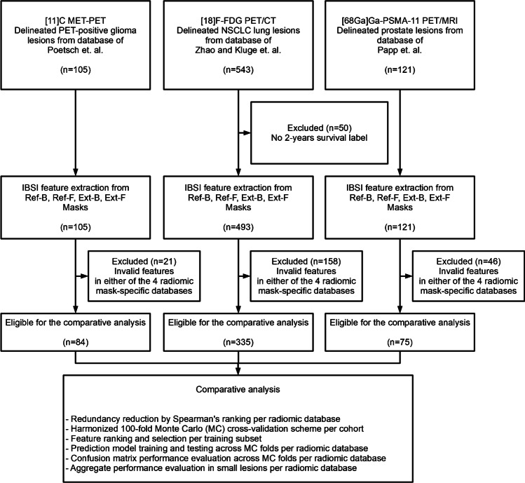
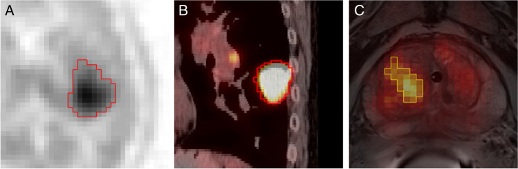
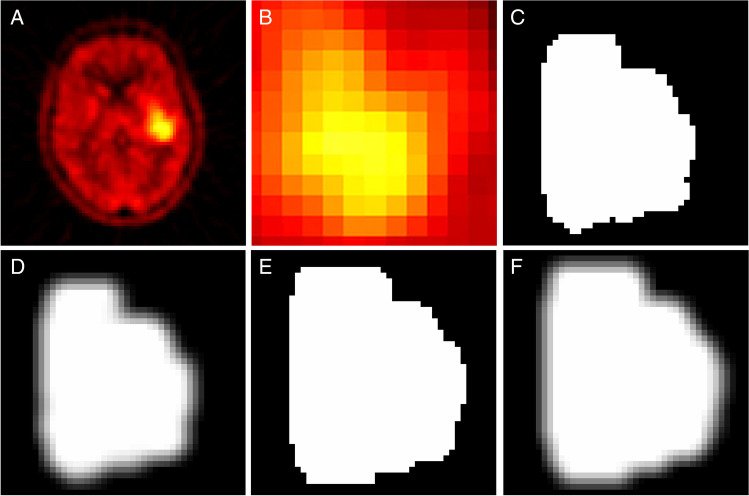
Previously published 11C Methionine (MET) positron emission tomography (PET) glioma, 18F-FDG PET/computed tomography (CT) lung, and 68GA-PSMA-11 PET/magneto-resonance imaging (MRI) prostate cancer retrospective cohorts were included in the analysis to predict their respective clinical endpoints. Four delineation methods including manually defined reference binary (Ref-B), its smoothed, fuzzified version (Ref-F), as well as extended binary (Ext-B) and its fuzzified version (Ext-F) were incorporated to extract imaging biomarker standardization initiative (IBSI)-conform radiomic features from each cohort. Machine learning for the four delineation approaches was performed utilizing a Monte Carlo cross-validation scheme to estimate the predictive performance of the four delineation methods.
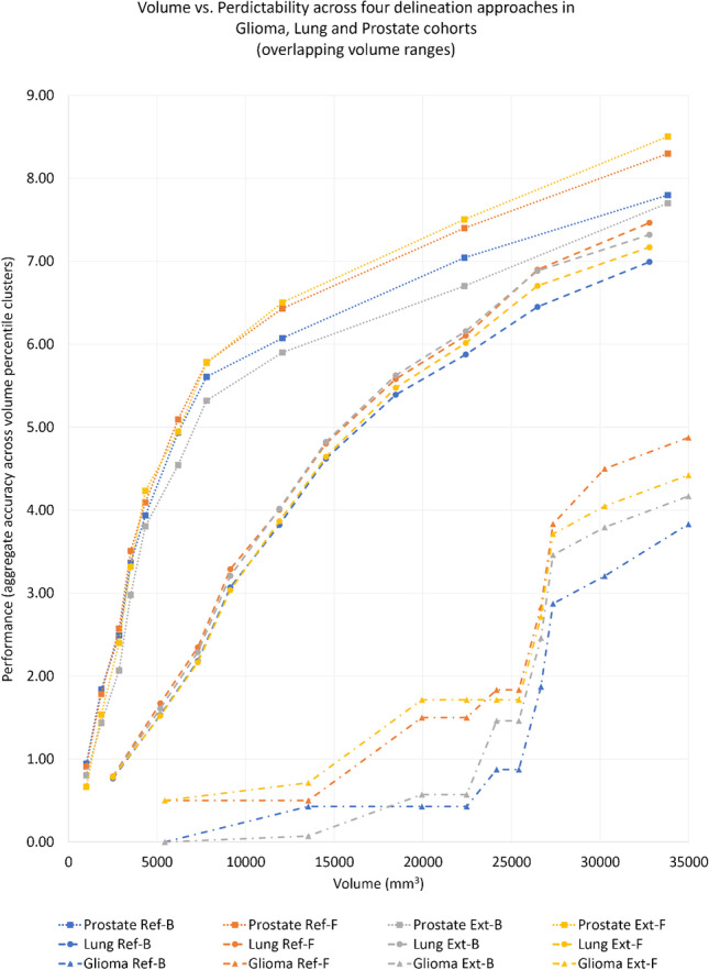
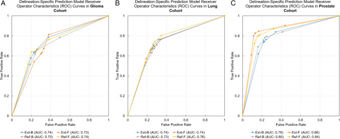
Reference fuzzy (Ref-F) delineation outperformed its binary delineation (Ref-B) counterpart in all cohorts within a volume range of 938-354987 mm with relative cross-validation area under the receiver operator characteristics curve (AUC) of +4.7-10.4. Compared to Ref-B, the highest AUC performance difference was observed by the Ref-F delineation in the glioma cohort (Ref-F: 0.74 vs. Ref-B: 0.70) and in the prostate cohort by Ref-F and Ext-F (Ref-F: 0.84, Ext-F: 0.86 vs. Ref-B: 0.80). In addition, fuzzy radiomics decreased feature redundancy by approx. 20%.
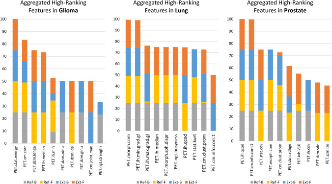
Fuzzy radiomics has the potential to increase predictive performance particularly in small lesion sizes compared to conventional binary radiomics in PET. We hypothesize that this effect is due to the ability of fuzzy radiomics to model partial volume effects and delineation uncertainties at small lesion boundaries. In addition, we consider that the lower redundancy of fuzzy radiomic features supports the identification of imaging biomarkers in future studies. Future studies shall consider systematically analyzing lesions and their surroundings with fuzzy and binary radiomics.
混合成像已成为医学成像的重要组成部分,特别是在临床常规中的癌症成像过程中。迄今为止,已有多项放射组学和机器学习研究调查了使用不同结果进行体内肿瘤特征描述的可行性。本研究旨在调查最近提出的模糊放射组学的效果,并将其预测性能与癌症成像队列中的常规放射组学进行比较。此外,还进行了病变与病变周围模糊和常规放射组学分析。
本研究纳入了先前发表的 11C 蛋氨酸(MET)正电子发射断层扫描(PET)脑胶质瘤、18F-FDG PET/计算机断层扫描(CT)肺癌和 68GA-PSMA-11 PET/磁共振成像(MRI)前列腺癌回顾性队列,以预测各自的临床终点。分析中纳入了四种勾画方法,包括手动定义的参考二进制(Ref-B)、其平滑、模糊版本(Ref-F)以及扩展二进制(Ext-B)及其模糊版本(Ext-F),以从每个队列中提取成像生物标志物标准化倡议(IBSI)一致的放射组学特征。对这四种勾画方法进行了机器学习,使用蒙特卡罗交叉验证方案来估计这四种勾画方法的预测性能。
在体积范围为 938-354987mm 的所有队列中,参考模糊(Ref-F)勾画均优于其二进制勾画(Ref-B),相对交叉验证受试者工作特征曲线(AUC)的 AUC 增加了 4.7-10.4。与 Ref-B 相比,Ref-F 勾画在脑胶质瘤队列中(Ref-F:0.74 比 Ref-B:0.70)和前列腺癌队列中(Ref-F 和 Ext-F:0.74、Ext-F:0.86 比 Ref-B:0.80)的 AUC 性能差异最大。此外,模糊放射组学降低了特征冗余度约 20%。
与传统的二进制放射组学相比,模糊放射组学具有在小病变大小的情况下提高预测性能的潜力,特别是在 PET 中。我们假设这种效果是由于模糊放射组学能够模拟小病变边界的部分容积效应和勾画不确定性。此外,我们认为模糊放射组学特征的较低冗余性支持在未来研究中识别成像生物标志物。未来的研究应考虑使用模糊和二进制放射组学系统地分析病变及其周围。