Department of Neurology, School of Medicine, Second Affiliated Hospital, Zhejiang University, Hangzhou, 310009, Zhejiang, China.
Department of Anesthesiology, School of Medicine, Women's Hospital, Zhejiang University, Hangzhou, 310009, Zhejiang, China.
J Neuroinflammation. 2023 Feb 5;20(1):26. doi: 10.1186/s12974-023-02709-w.
Inflammasome activation has a pathogenic role in Parkinson's disease (PD). Up-regulated expressions of inflammasome adaptor apoptosis-associated speck-like protein containing a CARD (ASC) and assembly of ASC specks have been observed in postmortems of human PD brains and experimental PD models. Extracellular ASC specks behave like danger signals and sustain prolonged inflammasome activation. However, the contribution of ASC specks in propagation of inflammasome activation and pathological progression in PD has not been fully established.
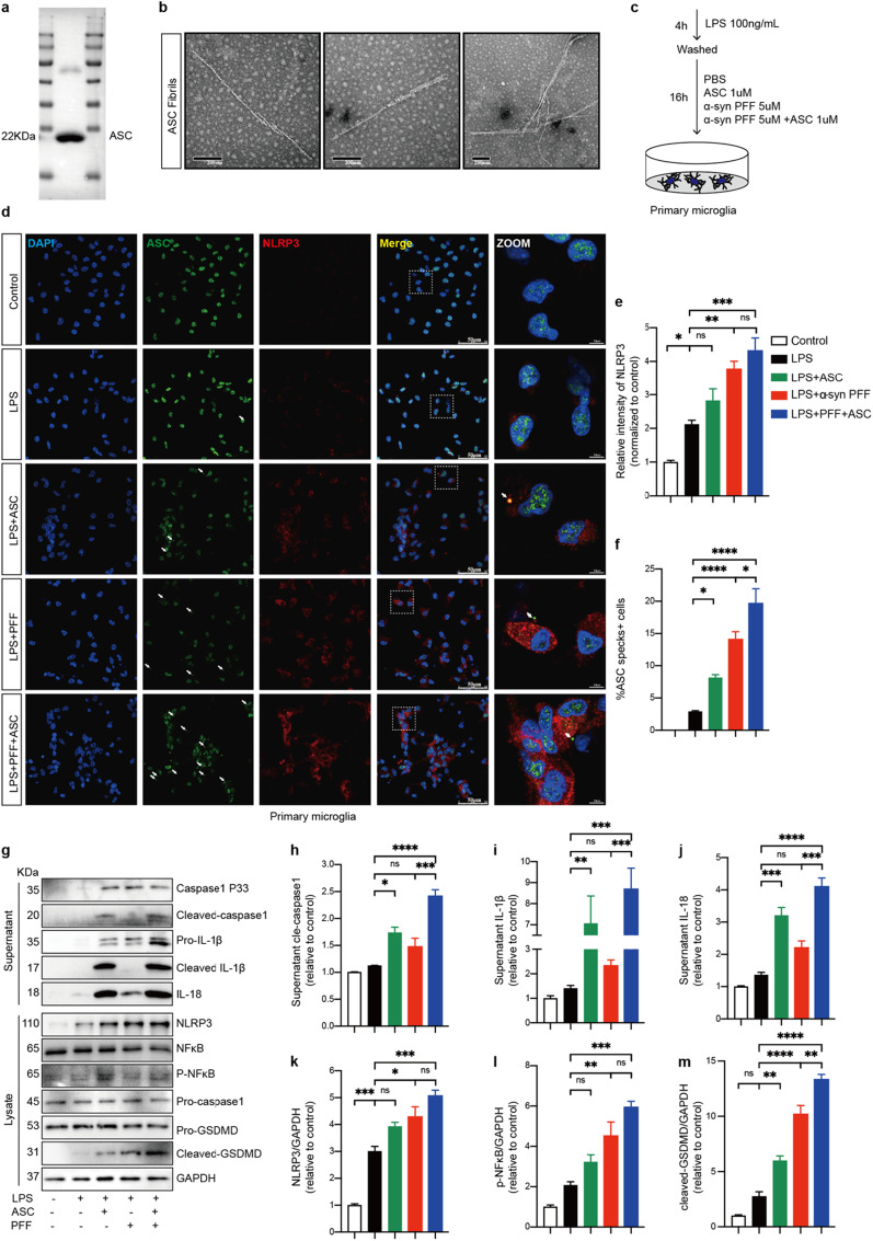
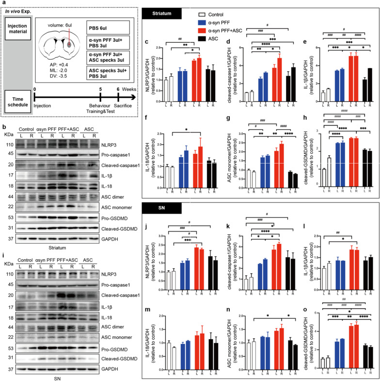
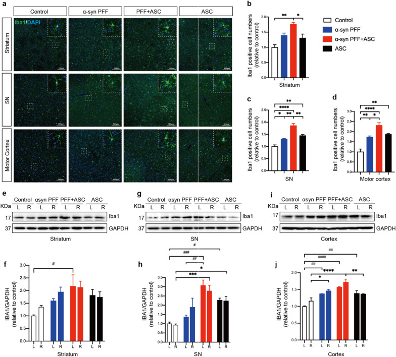
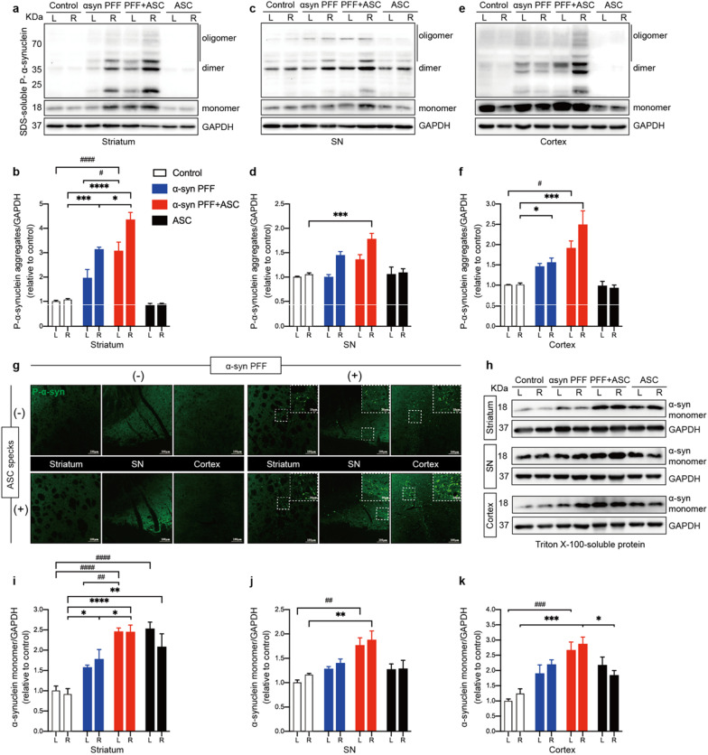
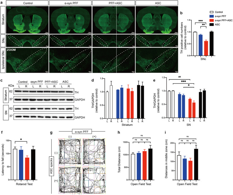
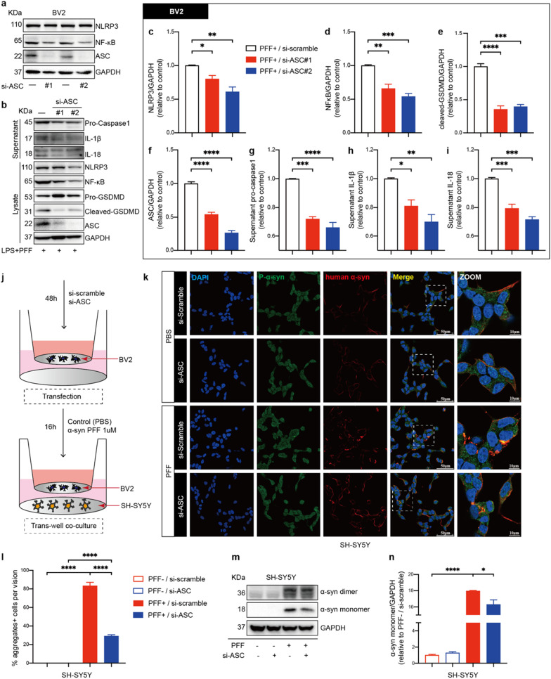
Herein, we used human A53T mutant α-synuclein preformed fibrils (PFFs)-stimulated microglia in vitro and unilateral striatal stereotaxic injection of PFFs-induced mice model of PD in vivo, to investigate the significance of ASC specks in PD pathological progression. Rotarod and open-field tests were performed to measure motor behaviors of indicated mice. Changes in the molecular expression were evaluated by immunofluorescence and immunoblotting (IB). Intracellular knockdown of the ASC in BV2 cells was performed using si-RNA. Microglial and neuronal cells were co-cultured in a trans-well system to determine the effects of ASC knockdown on cytoprotection.
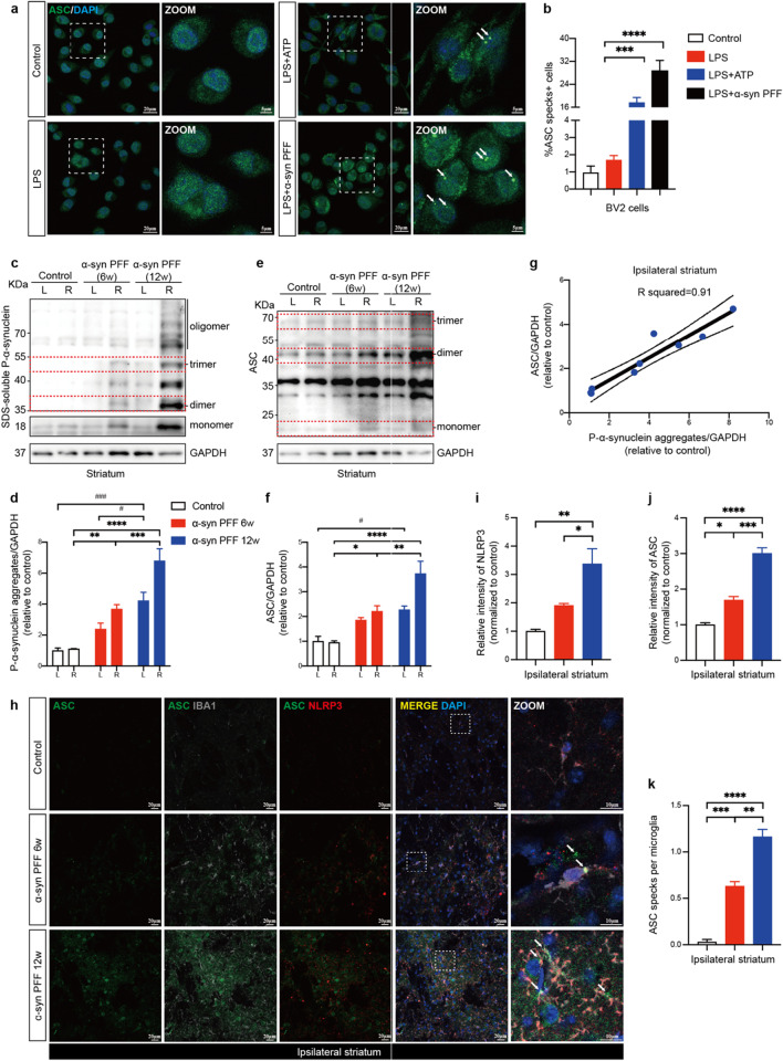
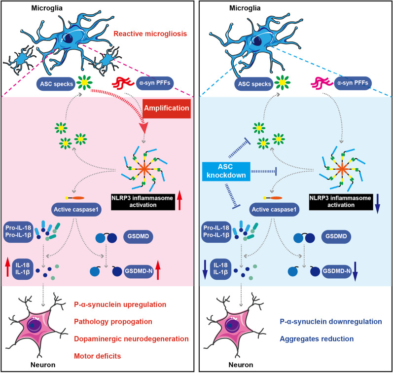
We observed a direct relationship between levels of ASC protein and misfolded α‑synuclein aggregates in PD mice brains. ASC specks amplified NLRP3 inflammasome activation driven by α-synuclein PFFs stimulation, which aggravated reactive microgliosis and accelerated α‑synuclein pathology, dopaminergic neurodegeneration and motor deficits. Endogenous ASC knockdown suppressed microglial inflammasome activation and neuronal α‑synuclein aggregation.
In conclusion, our study elucidated that ASC specks contribute to the propagation of inflammasome activation-associated α‑synuclein pathology in PD, which forms the basis for targeting ASC as a potential therapy for PD.
炎性小体的激活在帕金森病(PD)中具有致病作用。在人类 PD 大脑的尸检和实验性 PD 模型中,已观察到炎性小体衔接子凋亡相关斑点样蛋白(ASC)的上调表达和 ASC 斑点的组装。细胞外 ASC 斑点表现为危险信号,并维持炎性小体的持续激活。然而,ASC 斑点在 PD 中炎性小体激活和病理进展中的作用尚未完全确定。
本文采用体外 A53T 突变 α-突触核蛋白原纤维(PFFs)刺激小胶质细胞和体内单侧纹状体立体定位注射 PFFs 诱导的 PD 小鼠模型,研究 ASC 斑点在 PD 病理进展中的意义。通过转棒和旷场测试来测量指示性小鼠的运动行为。通过免疫荧光和免疫印迹(IB)评估分子表达的变化。使用 si-RNA 在 BV2 细胞中进行 ASC 的细胞内敲低。在 Trans-well 系统中进行小胶质细胞和神经元共培养,以确定 ASC 敲低对细胞保护的影响。
我们观察到 PD 小鼠大脑中 ASC 蛋白水平与错误折叠的α-突触核蛋白聚集之间存在直接关系。ASC 斑点放大了由α-突触核蛋白 PFFs 刺激驱动的 NLRP3 炎性小体激活,加重了反应性小胶质细胞增生,并加速了α-突触核蛋白病理学、多巴胺能神经退行性变和运动缺陷。内源性 ASC 敲低抑制了小胶质细胞炎性小体激活和神经元α-突触核蛋白聚集。
总之,我们的研究阐明了 ASC 斑点有助于 PD 中与炎性小体激活相关的α-突触核蛋白病理学的传播,这为靶向 ASC 作为 PD 潜在治疗方法提供了依据。