Amrute Junedh M, Luo Xin, Penna Vinay, Bredemeyer Andrea, Yamawaki Tracy, Yang Steven, Kadyrov Farid, Heo Gyu-Seong, Shi Sally Yu, Lee Paul, Koenig Andrew L, Kuppe Christoph, Jones Cameran, Kopecky Benjamin, Hayat Sikander, Ma Pan, Terada Yuriko, Fu Angela, Furtado Milena, Kreisel Daniel, Stitziel Nathan O, Li Chi-Ming, Kramann Rafael, Liu Yongjian, Ason Brandon, Lavine Kory J
Center for Cardiovascular Research, Division of Cardiology, Department of Medicine, Washington University School of Medicine, Saint Louis, MO, 63110, USA.
Genome Analysis Unit, Amgen Discovery Research, Amgen Inc., 1120 Veterans Blvd, South San Francisco, CA, 94080, USA.
Res Sq. 2023 Jan 26:rs.3.rs-2402606. doi: 10.21203/rs.3.rs-2402606/v1.
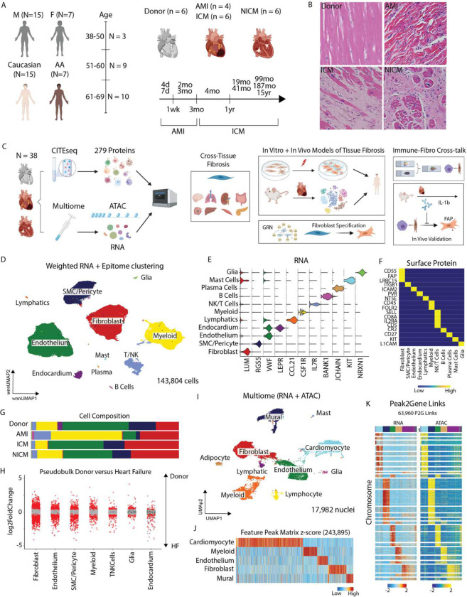
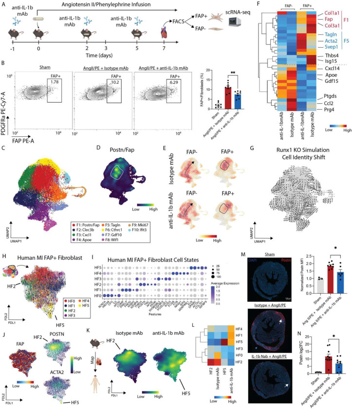
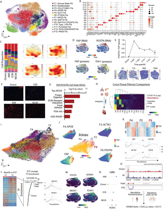
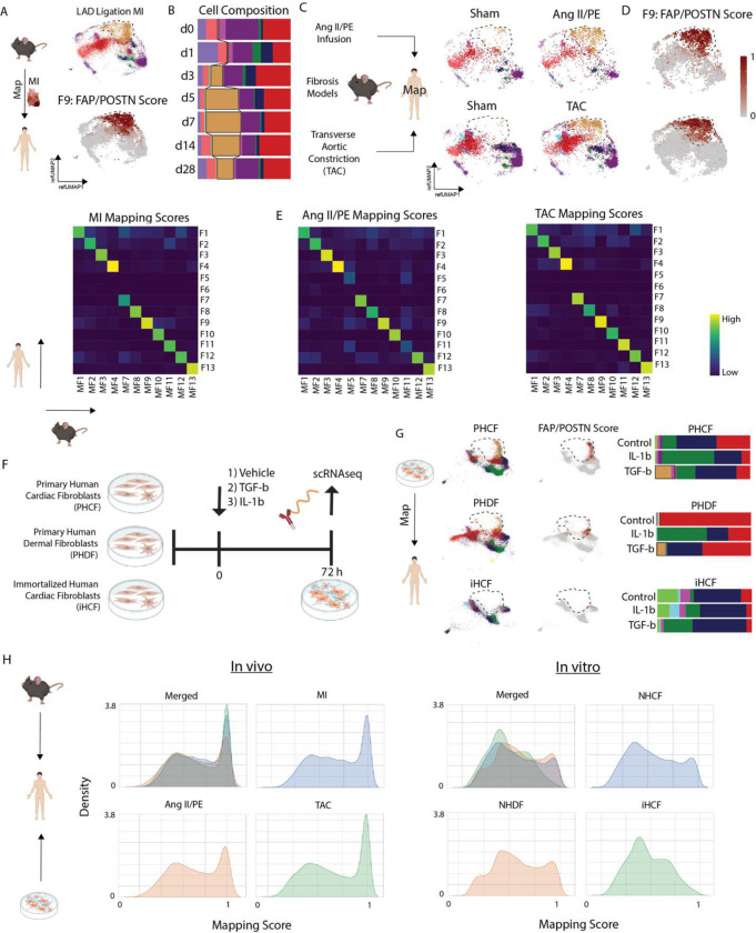
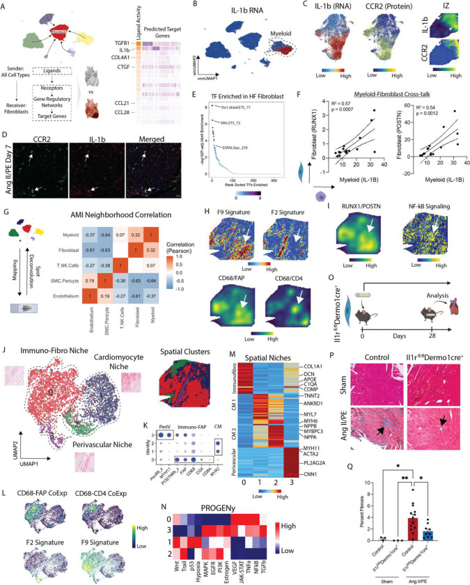
Inflammation and tissue fibrosis co-exist and are causally linked to organ dysfunction. However, the molecular mechanisms driving immune-fibroblast crosstalk in human cardiac disease remains unexplored and there are currently no therapeutics to target fibrosis. Here, we performed multi-omic single-cell gene expression, epitope mapping, and chromatin accessibility profiling in 38 donors, acutely infarcted, and chronically failing human hearts. We identified a disease-associated fibroblast trajectory marked by cell surface expression of fibroblast activator protein (FAP), which diverged into distinct myofibroblasts and pro-fibrotic fibroblast populations, the latter resembling matrifibrocytes. Pro-fibrotic fibroblasts were transcriptionally similar to cancer associated fibroblasts and expressed high levels of collagens and periostin (), thymocyte differentiation antigen 1 (THY-1), and endothelin receptor A () predicted to be driven by a gene regulatory network. We assessed the applicability of experimental systems to model tissue fibrosis and demonstrated that 3 different mouse models of cardiac injury were superior compared to cultured human heart and dermal fibroblasts in recapitulating the human disease phenotype. Ligand-receptor analysis and spatial transcriptomics predicted that interactions between C-C chemokine receptor type 2 (CCR2) macrophages and fibroblasts mediated by interleukin 1 beta (IL-1β) signaling drove the emergence of pro-fibrotic fibroblasts within spatially defined niches. This concept was validated through transcription factor perturbation and inhibition of IL-1β signaling in fibroblasts where we observed reduced pro-fibrotic fibroblasts, preferential differentiation of fibroblasts towards myofibroblasts, and reduced cardiac fibrosis. Herein, we show a subset of macrophages signal to fibroblasts via IL-1β and rewire their gene regulatory network and differentiation trajectory towards a pro-fibrotic fibroblast phenotype. These findings highlight the broader therapeutic potential of targeting inflammation to treat tissue fibrosis and restore organ function.
炎症与组织纤维化并存,且与器官功能障碍存在因果关系。然而,人类心脏疾病中驱动免疫细胞与成纤维细胞相互作用的分子机制仍未被探索,目前也没有针对纤维化的治疗方法。在此,我们对38名急性梗死和慢性衰竭的人类心脏供体进行了多组学单细胞基因表达、表位映射和染色质可及性分析。我们鉴定出一种与疾病相关的成纤维细胞轨迹,其特征是成纤维细胞激活蛋白(FAP)的细胞表面表达,该轨迹分化为不同的肌成纤维细胞和促纤维化成纤维细胞群体,后者类似于基质成纤维细胞。促纤维化成纤维细胞在转录上与癌症相关成纤维细胞相似,表达高水平的胶原蛋白和骨膜蛋白()、胸腺细胞分化抗原1(THY-1)以及内皮素受体A(),预计这些都是由一个基因调控网络驱动的。我们评估了实验系统模拟组织纤维化的适用性,并证明3种不同的心脏损伤小鼠模型在重现人类疾病表型方面优于培养的人类心脏和皮肤成纤维细胞。配体-受体分析和空间转录组学预测,由白细胞介素1β(IL-1β)信号介导的C-C趋化因子受体2(CCR2)巨噬细胞与成纤维细胞之间的相互作用,驱动了空间限定微环境中促纤维化成纤维细胞的出现。这一概念通过转录因子扰动和成纤维细胞中IL-1β信号的抑制得到验证,我们观察到促纤维化成纤维细胞减少、成纤维细胞向肌成纤维细胞的优先分化以及心脏纤维化减轻。在此,我们展示了一部分巨噬细胞通过IL-1β向成纤维细胞发出信号,并重新连接其基因调控网络和分化轨迹,使其向促纤维化成纤维细胞表型发展。这些发现突出了靶向炎症治疗组织纤维化和恢复器官功能的更广泛治疗潜力。