Department of Periodontology, College of Dentistry, University of Florida, Gainesville, FL 32610, USA.
Department of Nutrition and Food Science, University of Maryland, College Park, MD 20742, USA.
Int J Mol Sci. 2023 Jan 24;24(3):2327. doi: 10.3390/ijms24032327.
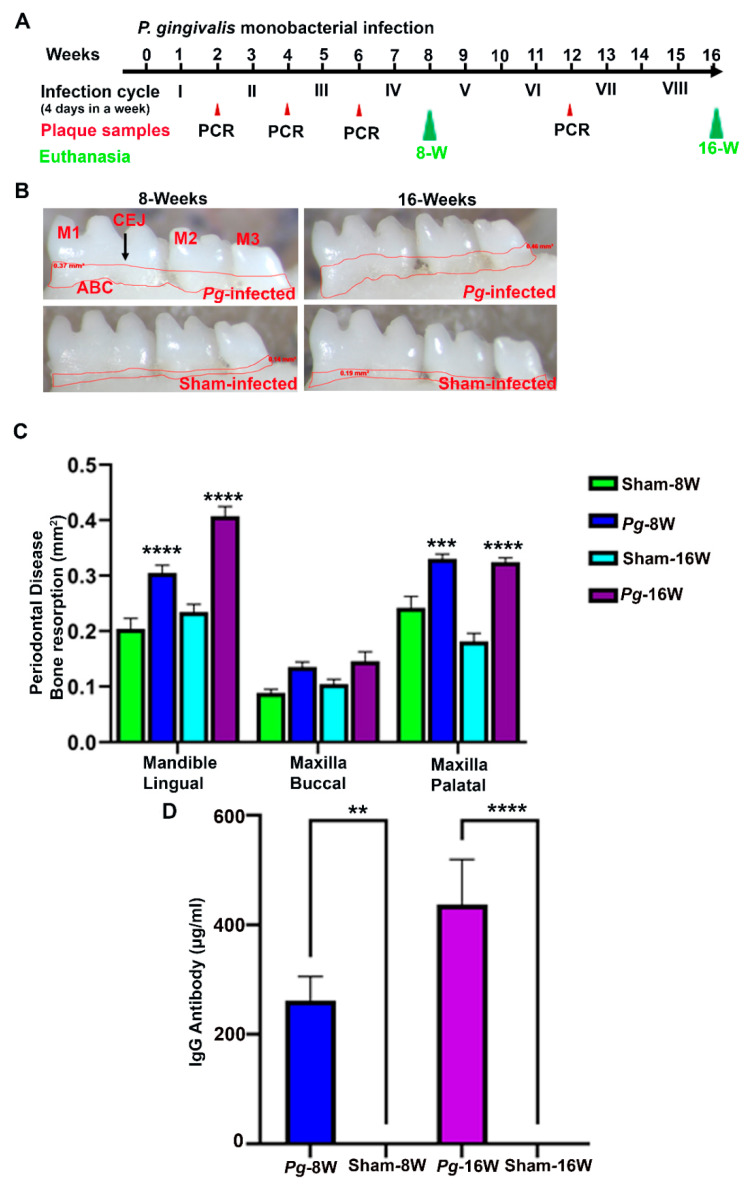
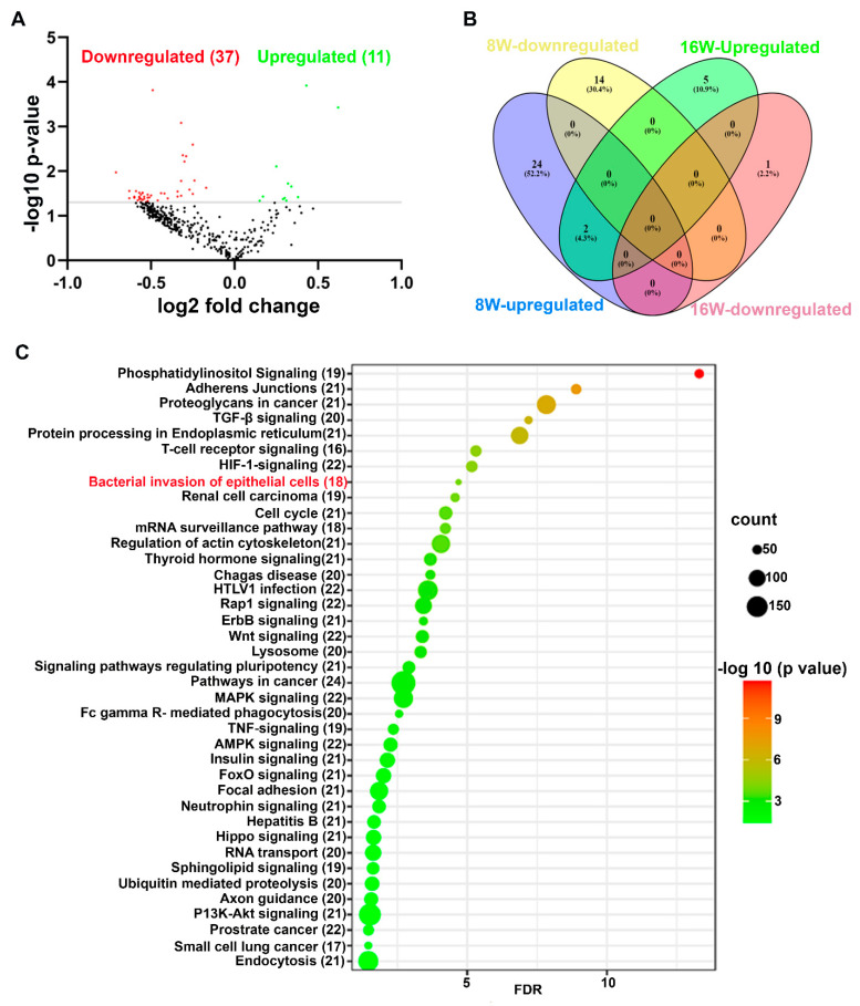
is one of the major bacteria constituting the subgingival pathogenic polymicrobial milieu during periodontitis. Our objective is to determine the global microRNA (miRNA, miR) expression kinetics in 8- and 16-weeks duration of infection in C57BL/6J mice and to identify the miRNA signatures at specific time-points in mice. We evaluated differential expression (DE) miRNAs in mandibles ( = 10) using high-throughput NanoString nCounter miRNA expression panels. The bacterial colonization, alveolar bone resorption (ABR), serum immunoglobulin G (IgG) antibodies, and bacterial dissemination were confirmed. In addition, all the infected mice showed bacterial colonization on the gingival surface, significant increases in ABR ( < 0.0001), and specific IgG antibody responses ( < 0.05-0.001). The miRNA profiling showed 26 upregulated miRNAs (e.g., miR-804, miR-690) and 14 downregulated miRNAs (e.g., miR-1902, miR-1937a) during an 8-weeks infection, whereas 7 upregulated miRNAs (e.g., miR-145, miR-195) and one downregulated miR-302b were identified during a 16-weeks infection. Both miR-103 and miR-30d were commonly upregulated at both time-points, and all the DE miRNAs were unique to the specific time-points. However, miR-31, miR-125b, miR-15a, and miR-195 observed in -infected mouse mandibles were also identified in the gingival tissues of periodontitis patients. None of the previously identified miRNAs reported in in vitro studies using cell lines (periodontal ligament cells, gingival epithelial cells, human leukemia monocytic cell line (THP-1), and B cells) exposed to lipopolysaccharide were observed in the in vivo study. Most of the pathways (endocytosis, bacterial invasion, and FcR-mediated phagocytosis) targeted by the DE miRNAs were linked with bacterial pathogen recognition and clearance. Further, eighteen miRNAs were closely associated with the bacterial invasion of epithelial cells. This study highlights the altered expression of miRNA in gingiva, and their expression depends on the time-points of infection. This is the first in vivo study that identified specific signature miRNAs (miR-103 and miR-30d) in invasion of epithelial cells, establishes a link between miRNA and development of periodontitis and helping to better understand the pathobiology of periodontitis.
是牙周炎患者龈下致病性多微生物环境中的主要细菌之一。我们的目的是确定在 8 周和 16 周的感染过程中 C57BL/6J 小鼠的全局 microRNA (miRNA,miR) 表达动力学,并确定在特定时间点在小鼠中的 miRNA 特征。我们使用高通量 NanoString nCounter miRNA 表达面板评估下颌骨 ( = 10) 中差异表达 (DE) 的 miRNA。细菌定植、牙槽骨吸收 (ABR)、血清免疫球蛋白 G (IgG) 抗体和细菌播散得到证实。此外,所有感染的小鼠在牙龈表面均有细菌定植, ABR 显著增加(<0.0001),特异性 IgG 抗体反应(<0.05-0.001)。miRNA 分析显示,在 8 周感染期间有 26 个上调的 miRNA(例如 miR-804、miR-690)和 14 个下调的 miRNA(例如 miR-1902、miR-1937a),而在 16 周感染期间有 7 个上调的 miRNA(例如 miR-145、miR-195)和一个下调的 miR-302b。miR-103 和 miR-30d 在两个时间点均共同上调,所有 DE miRNA 均为特定时间点所特有。然而,在感染的小鼠下颌骨中观察到的 miR-31、miR-125b、miR-15a 和 miR-195 也在牙周炎患者的牙龈组织中被鉴定出来。在体外使用细胞系(牙周韧带细胞、牙龈上皮细胞、人白血病单核细胞系 (THP-1) 和 B 细胞)暴露于脂多糖的情况下,在体内研究中未观察到先前在体外研究中报道的任何 DE miRNA。大多数 DE miRNA 靶向的途径(内吞作用、细菌入侵和 FcR 介导的吞噬作用)与细菌病原体的识别和清除有关。此外,有 18 个 miRNA 与上皮细胞的细菌入侵密切相关。本研究强调了 miRNA 在牙龈中的表达改变,并且它们的表达取决于感染的时间点。这是第一项在体内研究中鉴定出特定的上皮细胞入侵的特征性 miRNA(miR-103 和 miR-30d)的研究,建立了 miRNA 与牙周炎发生发展之间的联系,并有助于更好地理解牙周炎的病理生物学。